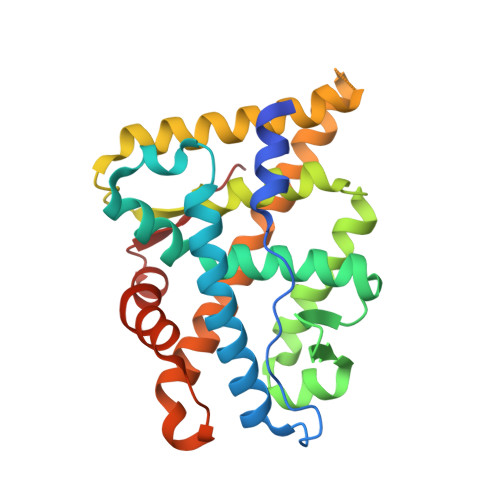
A partially open conformation of an androgen receptor ligand-binding domain with drug-resistance mutations.
Doamekpor, S.K., Peng, P., Xu, R., Ma, L., Tong, Y., Tong, L.(2023) Acta Crystallogr F Struct Biol Commun 79: 95-104
- PubMed: 36995121 Search on PubMedSearch on PubMed Central
- DOI: https://doi.org/10.1107/S2053230X23002224
- Primary Citation Related Structures:
8FGY, 8FGZ, 8FH0, 8FH1, 8FH2 - PubMed Abstract:
Mutations in the androgen receptor (AR) ligand-binding domain (LBD) can cause resistance to drugs used to treat prostate cancer. Commonly found mutations include L702H, W742C, H875Y, F877L and T878A, while the F877L mutation can convert second-generation antagonists such as enzalutamide and apalutamide into agonists. However, pruxelutamide, another second-generation AR antagonist, has no agonist activity with the F877L and F877L/T878A mutants and instead maintains its inhibitory activity against them. Here, it is shown that the quadruple mutation L702H/H875Y/F877L/T878A increases the soluble expression of AR LBD in complex with pruxelutamide in Escherichia coli. The crystal structure of the quadruple mutant in complex with the agonist dihydrotestosterone (DHT) reveals a partially open conformation of the AR LBD due to conformational changes in the loop connecting helices H11 and H12 (the H11-H12 loop) and Leu881. This partially open conformation creates a larger ligand-binding site for AR. Additional structural studies suggest that both the L702H and F877L mutations are important for conformational changes. This structural variability in the AR LBD could affect ligand binding as well as the resistance to antagonists.
- Department of Biological Sciences, Columbia University, New York, NY 10027, USA.
Organizational Affiliation: