8FFX | pdb_00008ffx

Crystal structure of HIV-1 reverse transcriptase in complex with non-nucleoside inhibitor 19980


Experimental Data Snapshot

  • Method: X-RAY DIFFRACTION
  • Resolution: 2.42 Å
  • R-Value Free: 
    0.266 (Depositor), 0.266 (DCC) 
  • R-Value Work: 
    0.214 (Depositor), 0.214 (DCC) 
  • R-Value Observed: 
    0.216 (Depositor) 

Starting Model: experimental
View more details

wwPDB Validation 3D Report Full Report

Validation slider image for 8FFX

Ligand Structure Quality Assessment 


This is version 1.1 of the entry. See complete history

Literature

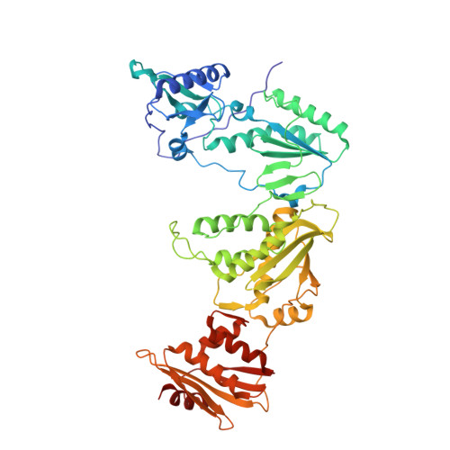
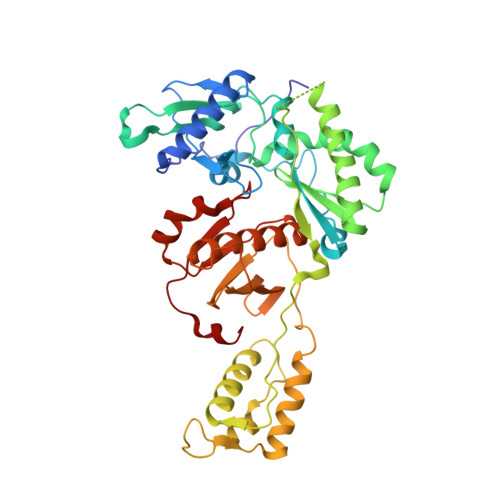
Targeting HIV-1 Reverse Transcriptase Using a Fragment-Based Approach.

Mansouri, M.Rumrill, S.Dawson, S.Johnson, A.Pinson, J.A.Gunzburg, M.J.Latham, C.F.Barlow, N.Mbogo, G.W.Ellenberg, P.Headey, S.J.Sluis-Cremer, N.Tyssen, D.Bauman, J.D.Ruiz, F.X.Arnold, E.Chalmers, D.K.Tachedjian, G.

(2023) Molecules 28

  • DOI: https://doi.org/10.3390/molecules28073103
  • Primary Citation Related Structures: 
    8FFX

  • PubMed Abstract: 

    Human immunodeficiency virus type I (HIV-1) is a retrovirus that infects cells of the host's immune system leading to acquired immunodeficiency syndrome and potentially death. Although treatments are available to prevent its progression, HIV-1 remains a major burden on health resources worldwide. Continued emergence of drug-resistance mutations drives the need for novel drugs that can inhibit HIV-1 replication through new pathways. The viral protein reverse transcriptase (RT) plays a fundamental role in the HIV-1 replication cycle, and multiple approved medications target this enzyme. In this study, fragment-based drug discovery was used to optimize a previously identified hit fragment (compound B-1 ), which bound RT at a novel site. Three series of compounds were synthesized and evaluated for their HIV-1 RT binding and inhibition. These series were designed to investigate different vectors around the initial hit in an attempt to improve inhibitory activity against RT. Our results show that the 4-position of the core scaffold is important for binding of the fragment to RT, and a lead compound with a cyclopropyl substitution was selected and further investigated. Requirements for binding to the NNRTI-binding pocket (NNIBP) and a novel adjacent site were investigated, with lead compound 27 -a minimal but efficient NNRTI-offering a starting site for the development of novel dual NNIBP-Adjacent site inhibitors.


  • Organizational Affiliation
    • Medicinal Chemistry, Monash Institute of Pharmaceutical Sciences, Monash University, Parkville, VIC 3052, Australia.

Macromolecule Content 

  • Total Structure Weight: 114.84 kDa 
  • Atom Count: 8,124 
  • Modeled Residue Count: 971 
  • Deposited Residue Count: 985 
  • Unique protein chains: 2

Macromolecules

Find similar proteins by:|  3D Structure
Entity ID: 1
MoleculeChains  Sequence LengthOrganismDetailsImage
Reverse transcriptase/ribonuclease H557Human immunodeficiency virus type 1 BH10Mutation(s): 3 
Gene Names: gag-pol
EC: 2.7.7.49 (PDB Primary Data), 2.7.7.7 (PDB Primary Data), 3.1.26.13 (PDB Primary Data), 3.1.13.2 (PDB Primary Data)
UniProt
Find proteins for P03366 (Human immunodeficiency virus type 1 group M subtype B (isolate BH10))
Explore P03366 
Go to UniProtKB:  P03366
Entity Groups
Sequence Clusters30% Identity50% Identity70% Identity90% Identity95% Identity100% Identity
UniProt GroupP03366
Sequence Annotations
Expand
Reference Sequence
Find similar proteins by:|  3D Structure
Entity ID: 2
MoleculeChains  Sequence LengthOrganismDetailsImage
p51 RT428Human immunodeficiency virus type 1 BH10Mutation(s): 1 
Gene Names: gag-pol
UniProt
Find proteins for P03366 (Human immunodeficiency virus type 1 group M subtype B (isolate BH10))
Explore P03366 
Go to UniProtKB:  P03366
Entity Groups
Sequence Clusters30% Identity50% Identity70% Identity90% Identity95% Identity100% Identity
UniProt GroupP03366
Sequence Annotations
Expand
Reference Sequence

Small Molecules

Ligands 4 Unique
IDChains Name / Formula / InChI Key2D Diagram3D Interactions
XU9
(Subject of Investigation/LOI)

Query on XU9


Download:Ideal Coordinates CCD File
D [auth A]ethyl (8S)-4-(cyclopropylethynyl)pyrazolo[1,5-a]pyridine-3-carboxylate
C15 H14 N2 O2
JXKYSMBFBAAONQ-UHFFFAOYSA-N
SO4

Query on SO4


Download:Ideal Coordinates CCD File
F [auth A],
G [auth A],
J [auth B]
SULFATE ION
O4 S
QAOWNCQODCNURD-UHFFFAOYSA-L
DMS

Query on DMS


Download:Ideal Coordinates CCD File
C [auth A]DIMETHYL SULFOXIDE
C2 H6 O S
IAZDPXIOMUYVGZ-UHFFFAOYSA-N
EDO

Query on EDO


Download:Ideal Coordinates CCD File
E [auth A],
H [auth B],
I [auth B]
1,2-ETHANEDIOL
C2 H6 O2
LYCAIKOWRPUZTN-UHFFFAOYSA-N

Experimental Data & Validation

Experimental Data

  • Method: X-RAY DIFFRACTION
  • Resolution: 2.42 Å
  • R-Value Free:  0.266 (Depositor), 0.266 (DCC) 
  • R-Value Work:  0.214 (Depositor), 0.214 (DCC) 
  • R-Value Observed: 0.216 (Depositor) 
Space Group: C 1 2 1
Unit Cell:
Length ( Å )Angle ( ˚ )
a = 161.952α = 90
b = 73.377β = 100.05
c = 107.966γ = 90
Software Package:
Software NamePurpose
PHENIXrefinement
autoXDSdata reduction
pointlessdata scaling
PHASERphasing

Structure Validation

View Full Validation Report


Ligand Structure Quality Assessment 


Entry History 

& Funding Information

Deposition Data


Funding OrganizationLocationGrant Number
National Institutes of Health/National Institute Of Allergy and Infectious Diseases (NIH/NIAID)United States--

Revision History  (Full details and data files)

  • Version 1.0: 2023-04-26
    Type: Initial release
  • Version 1.1: 2024-05-22
    Changes: Data collection