


CryoEM structure of bacteriophage Q-beta coat protein dimer with AYGG linker
Newton, T.P., Zhao, L., Kopylov, M., Finn, M.G.To be published.
Experimental Data Snapshot
wwPDB Validation 3D Report Full Report

Entity ID: 1 | |||||
|---|---|---|---|---|---|
| Molecule | Chains | Sequence Length | Organism | Details | Image |
| Minor capsid protein A1 fusion | A [auth D] | 268 | Qubevirus durum | Mutation(s): 0 | 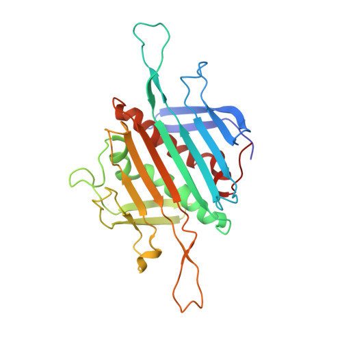 |
UniProt | |||||
Entity Groups | |||||
| Sequence Clusters | 30% Identity50% Identity70% Identity90% Identity95% Identity100% Identity | ||||
| UniProt Group | Q8LTE1 | ||||
Sequence AnnotationsExpand | |||||
Reference Sequence | |||||
| Funding Organization | Location | Grant Number |
|---|---|---|
| Simons Foundation | United States | SF349247 |


