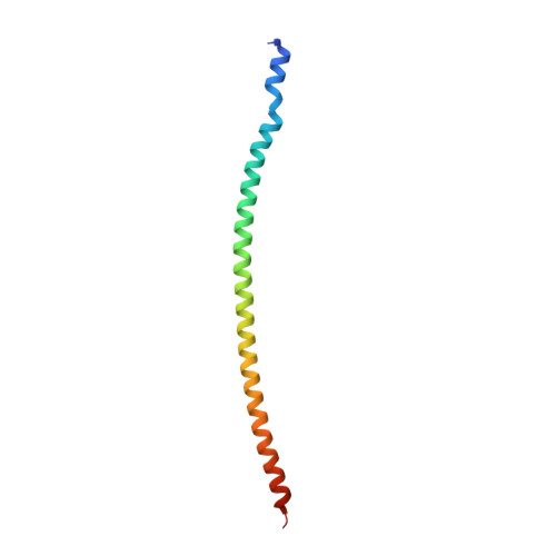
8FBY

Crystal structure of PLVAP CC1 determined by sulfur SAD

  • Classification: MEMBRANE PROTEIN
  • Organism(s): Mus musculus
  • Expression System: Escherichia coli
  • Mutation(s): No 

  • Deposited: 2022-11-30 Released: 2023-03-22 
  • Deposition Author(s): Chang, T.H., Hsieh, F.L., Gabelli, S.B., Nathans, J.
  • Funding Organization(s): National Institutes of Health/National Eye Institute (NIH/NEI), National Institutes of Health/National Cancer Institute (NIH/NCI), Human Frontier Science Program (HFSP), Howard Hughes Medical Institute (HHMI)

Experimental Data Snapshot

  • Method: X-RAY DIFFRACTION
  • Resolution: 2.40 Å
  • R-Value Free: 0.261 
  • R-Value Work: 0.223 
  • R-Value Observed: 0.225 

wwPDB Validation   3D Report Full Report


This is version 1.1 of the entry. See complete history


Literature

Structural insights into plasmalemma vesicle-associated protein (PLVAP): Implications for vascular endothelial diaphragms and fenestrae.

Chang, T.H.Hsieh, F.L.Gu, X.Smallwood, P.M.Kavran, J.M.Gabelli, S.B.Nathans, J.

(2023) Proc Natl Acad Sci U S A 120: e2221103120-e2221103120

  • DOI: https://doi.org/10.1073/pnas.2221103120
  • Primary Citation of Related Structures:  
    8FBY, 8FCF

  • PubMed Abstract: 

    In many organs, small openings across capillary endothelial cells (ECs) allow the diffusion of low-molecular weight compounds and small proteins between the blood and tissue spaces. These openings contain a diaphragm composed of radially arranged fibers, and current evidence suggests that a single-span type II transmembrane protein, plasmalemma vesicle-associated protein-1 (PLVAP), constitutes these fibers. Here, we present the three-dimensional crystal structure of an 89-amino acid segment of the PLVAP extracellular domain (ECD) and show that it adopts a parallel dimeric alpha-helical coiled-coil configuration with five interchain disulfide bonds. The structure was solved using single-wavelength anomalous diffraction from sulfur-containing residues (sulfur SAD) to generate phase information. Biochemical and circular dichroism (CD) experiments show that a second PLVAP ECD segment also has a parallel dimeric alpha-helical configuration-presumably a coiled coil-held together with interchain disulfide bonds. Overall, ~2/3 of the ~390 amino acids within the PLVAP ECD adopt a helical configuration, as determined by CD. We also determined the sequence and epitope of MECA-32, an anti-PLVAP antibody. Taken together, these data lend strong support to the model of capillary diaphragms formulated by Tse and Stan in which approximately ten PLVAP dimers are arranged within each 60- to 80-nm-diameter opening like the spokes of a bicycle wheel. Passage of molecules through the wedge-shaped pores is presumably determined both by the length of PLVAP-i.e., the long dimension of the pore-and by the chemical properties of amino acid side chains and N-linked glycans on the solvent-accessible faces of PLVAP.


  • Organizational Affiliation

    Department of Molecular Biology and Genetics, Johns Hopkins University School of Medicine, Baltimore, MD 21205.


Macromolecules
Find similar proteins by:  (by identity cutoff)  |  3D Structure
Entity ID: 1
MoleculeChains Sequence LengthOrganismDetailsImage
Plasmalemma vesicle-associated protein
A, B, C, D
95Mus musculusMutation(s): 0 
Gene Names: PlvapPv1
UniProt
Find proteins for Q91VC4 (Mus musculus)
Explore Q91VC4 
Go to UniProtKB:  Q91VC4
Entity Groups  
Sequence Clusters30% Identity50% Identity70% Identity90% Identity95% Identity100% Identity
UniProt GroupQ91VC4
Sequence Annotations
Expand
  • Reference Sequence
Experimental Data & Validation

Experimental Data

  • Method: X-RAY DIFFRACTION
  • Resolution: 2.40 Å
  • R-Value Free: 0.261 
  • R-Value Work: 0.223 
  • R-Value Observed: 0.225 
  • Space Group: P 21 21 21
Unit Cell:
Length ( Å )Angle ( ˚ )
a = 35.587α = 90
b = 86.232β = 90
c = 181.647γ = 90
Software Package:
Software NamePurpose
BUSTERrefinement
Aimlessdata scaling
xia2data reduction
AutoSolphasing

Structure Validation

View Full Validation Report


Entry History & Funding Information

Deposition Data


Funding OrganizationLocationGrant Number
National Institutes of Health/National Eye Institute (NIH/NEI)United States--
National Institutes of Health/National Cancer Institute (NIH/NCI)United States--
Human Frontier Science Program (HFSP)France--
Howard Hughes Medical Institute (HHMI)United States--

Revision History  (Full details and data files)

  • Version 1.0: 2023-03-22
    Type: Initial release
  • Version 1.1: 2023-04-12
    Changes: Database references