Structures of Mycobacterium tuberculosis isoprenyl diphosphate synthase Rv2173 in substrate-bound forms.
Titterington, J.A., Ho, N.A.T., Beasley, C.P.H., Mann, F., Baker, E.N., Allison, T.M., Johnston, J.M.(2025) Acta Crystallogr F Struct Biol Commun 81: 193-200
- PubMed: 40166974 Search on PubMedSearch on PubMed Central
- DOI: https://doi.org/10.1107/S2053230X25002298
- Primary Citation Related Structures:
8F8F, 8F8K, 8F8L - PubMed Abstract:
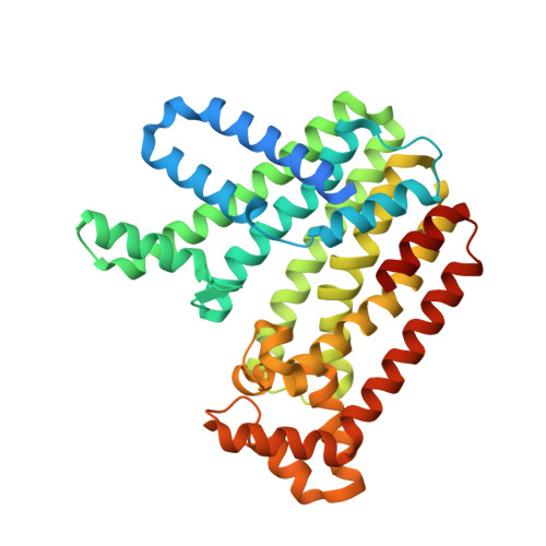
We report structures of the Mycobacterium tuberculosis isoprenyl diphosphate synthase Rv2173 in three forms: apo and two substrate-bound forms [isoprenyl diphosphate (IPP) and dimethylallyl diphosphate (DMAPP)]. The protein possesses a canonical all-α-helical trans-isoprenyl diphosphate synthase fold that is dimeric in each form. There are some differences between the structures: the IPP-bound form shows IPP bound in the DMAPP/allylic substrate-binding site with three divalent metal ions bound around the IPP and the complete C-terminus closing around the active site, while the apo and DMAPP-bound forms are more open, with some of the C-terminal region disordered, supporting suggestions that the C-terminus is important in substrate entry/product exit. In the DMAPP form DMAPP occupies the expected allylic substrate site, but only two metal ions are associated with the binding, with the DMAPP diphosphates adopting a slightly different binding pose compared with IPP in the same site, and the third metal-binding site is unoccupied. In no case is the IPP binding site occupied by IPP. There has been some uncertainty regarding product length for Rv2173, with variable lengths being reported. In the structures reported here, the `capping' residue at the bottom of the binding cavity is tryptophan and comparison with other IPP synthases suggests that the structure of Rv2173 is most consistent with a C 10 -C 15 final product size.
- Biomolecular Interaction Centre and School of Physical and Chemical Sciences, University of Canterbury. Christchurch, New Zealand.
Organizational Affiliation: