8F6D | pdb_00008f6d

Crystal structure of the CNNM2 CBS-pair domain in complex with ARL15


Experimental Data Snapshot

  • Method: X-RAY DIFFRACTION
  • Resolution: 3.20 Å
  • R-Value Free: 
    0.293 (Depositor), 0.292 (DCC) 
  • R-Value Work: 
    0.264 (Depositor), 0.264 (DCC) 
  • R-Value Observed: 
    0.266 (Depositor) 

Starting Model: experimental
View more details

wwPDB Validation 3D Report Full Report

Validation slider image for 8F6D

This is version 1.2 of the entry. See complete history

Literature

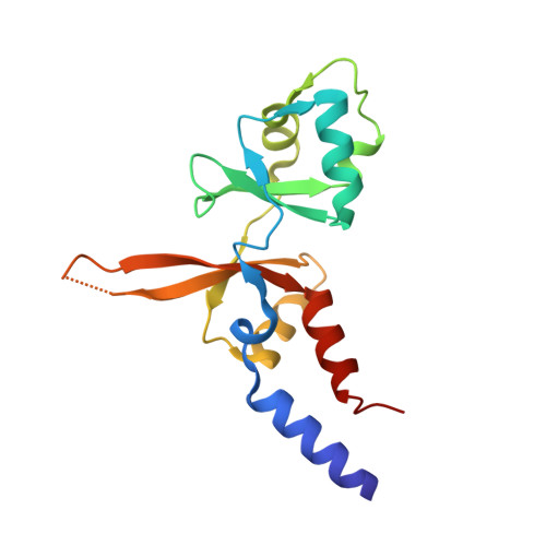
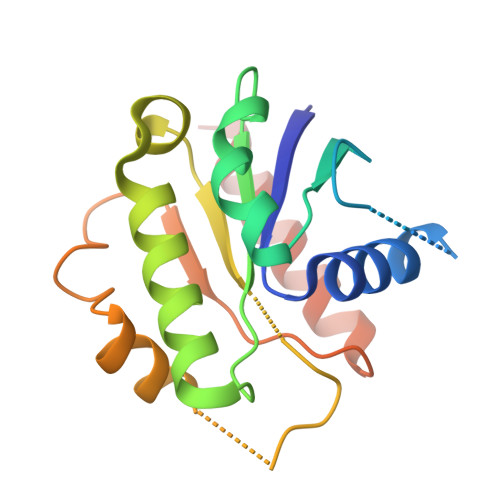
Structural insights into regulation of CNNM-TRPM7 divalent cation uptake by the small GTPase ARL15.

Mahbub, L.Kozlov, G.Zong, P.Lee, E.L.Tetteh, S.Nethramangalath, T.Knorn, C.Jiang, J.Shahsavan, A.Yue, L.Runnels, L.Gehring, K.

(2023) Elife 12

  • DOI: https://doi.org/10.7554/eLife.86129
  • Primary Citation Related Structures: 
    8F6D

  • PubMed Abstract: 

    Cystathionine-β-synthase (CBS)-pair domain divalent metal cation transport mediators (CNNMs) are an evolutionarily conserved family of magnesium transporters. They promote efflux of Mg 2+ ions on their own and influx of divalent cations when expressed with the transient receptor potential ion channel subfamily M member 7 (TRPM7). Recently, ADP-ribosylation factor-like GTPase 15 (ARL15) has been identified as CNNM-binding partner and an inhibitor of divalent cation influx by TRPM7. Here, we characterize ARL15 as a GTP and CNNM-binding protein and demonstrate that ARL15 also inhibits CNNM2 Mg 2+ efflux. The crystal structure of a complex between ARL15 and CNNM2 CBS-pair domain reveals the molecular basis for binding and allowed the identification of mutations that specifically block binding. A binding deficient ARL15 mutant, R95A, failed to inhibit CNNM and TRPM7 transport of Mg 2+ and Zn 2+ ions. Structural analysis and binding experiments with phosphatase of regenerating liver 2 (PRL2 or PTP4A2) showed that ARL15 and PRLs compete for binding CNNM to coordinate regulation of ion transport by CNNM and TRPM7.


  • Organizational Affiliation
    • Department of Biochemistry, McGill University, Montreal, Canada.

Macromolecule Content 

  • Total Structure Weight: 149.28 kDa 
  • Atom Count: 7,791 
  • Modeled Residue Count: 1,146 
  • Deposited Residue Count: 1,316 
  • Unique protein chains: 2

Macromolecules

Find similar proteins by:|  3D Structure
Entity ID: 1
MoleculeChains  Sequence LengthOrganismDetailsImage
Metal transporter CNNM2
A, C, E, G
156Homo sapiensMutation(s): 0 
Gene Names: CNNM2ACDP2
UniProt & NIH Common Fund Data Resources
Find proteins for Q9H8M5 (Homo sapiens)
Explore Q9H8M5 
Go to UniProtKB:  Q9H8M5
PHAROS:  Q9H8M5
GTEx:  ENSG00000148842 
Entity Groups
Sequence Clusters30% Identity50% Identity70% Identity90% Identity95% Identity100% Identity
UniProt GroupQ9H8M5
Sequence Annotations
Expand
Reference Sequence
Find similar proteins by:|  3D Structure
Entity ID: 2
MoleculeChains  Sequence LengthOrganismDetailsImage
ADP-ribosylation factor-like protein 15
B, D, F, H
173Homo sapiensMutation(s): 0 
Gene Names: ARL15ARFRP2
UniProt & NIH Common Fund Data Resources
Find proteins for Q9NXU5 (Homo sapiens)
Explore Q9NXU5 
Go to UniProtKB:  Q9NXU5
PHAROS:  Q9NXU5
GTEx:  ENSG00000185305 
Entity Groups
Sequence Clusters30% Identity50% Identity70% Identity90% Identity95% Identity100% Identity
UniProt GroupQ9NXU5
Sequence Annotations
Expand
Reference Sequence

Experimental Data & Validation

Experimental Data

  • Method: X-RAY DIFFRACTION
  • Resolution: 3.20 Å
  • R-Value Free:  0.293 (Depositor), 0.292 (DCC) 
  • R-Value Work:  0.264 (Depositor), 0.264 (DCC) 
  • R-Value Observed: 0.266 (Depositor) 
Space Group: P 1
Unit Cell:
Length ( Å )Angle ( ˚ )
a = 66.018α = 94.353
b = 73.323β = 95.5
c = 79.327γ = 115.33
Software Package:
Software NamePurpose
PHENIXrefinement
DIALSdata reduction
DIALSdata scaling
PHENIXphasing

Structure Validation

View Full Validation Report


Entry History 

& Funding Information

Deposition Data


Funding OrganizationLocationGrant Number
Canadian Institutes of Health Research (CIHR)Canada--

Revision History  (Full details and data files)

  • Version 1.0: 2023-07-12
    Type: Initial release
  • Version 1.1: 2023-10-25
    Changes: Data collection, Refinement description
  • Version 1.2: 2024-01-24
    Changes: Database references