8F5D | pdb_00008f5d

Architecture of the MurE-MurF ligase bacterial cell wall biosynthesis complex


Experimental Data Snapshot

  • Method: X-RAY DIFFRACTION
  • Resolution: 2.56 Å
  • R-Value Free: 
    0.292 (Depositor), 0.290 (DCC) 
  • R-Value Work: 
    0.249 (Depositor), 0.250 (DCC) 
  • R-Value Observed: 
    0.253 (Depositor) 

Starting Model: in silico
View more details

wwPDB Validation   3D Report Full Report


This is version 1.1 of the entry. See complete history


Literature

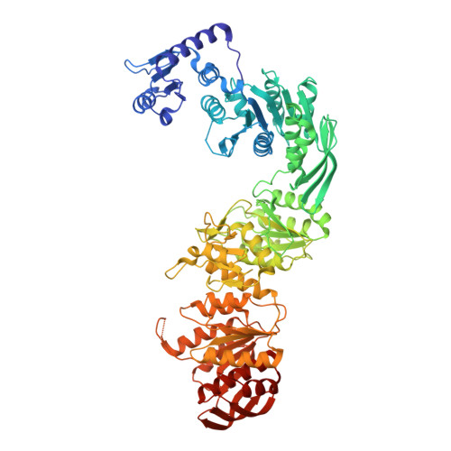
Architecture and genomic arrangement of the MurE-MurF bacterial cell wall biosynthesis complex.

Shirakawa, K.T.Sala, F.A.Miyachiro, M.M.Job, V.Trindade, D.M.Dessen, A.

(2023) Proc Natl Acad Sci U S A 120: e2219540120-e2219540120

  • DOI: https://doi.org/10.1073/pnas.2219540120
  • Primary Citation of Related Structures:  
    8F5D

  • PubMed Abstract: 

    Peptidoglycan (PG) is a central component of the bacterial cell wall, and the disruption of its biosynthetic pathway has been a successful antibacterial strategy for decades. PG biosynthesis is initiated in the cytoplasm through sequential reactions catalyzed by Mur enzymes that have been suggested to associate into a multimembered complex. This idea is supported by the observation that in many eubacteria, mur genes are present in a single operon within the well conserved dcw cluster, and in some cases, pairs of mur genes are fused to encode a single, chimeric polypeptide. We performed a vast genomic analysis using >140 bacterial genomes and mapped Mur chimeras in numerous phyla, with Proteobacteria carrying the highest number. MurE-MurF, the most prevalent chimera, exists in forms that are either directly associated or separated by a linker. The crystal structure of the MurE-MurF chimera from Bordetella pertussis reveals a head-to-tail, elongated architecture supported by an interconnecting hydrophobic patch that stabilizes the positions of the two proteins. Fluorescence polarization assays reveal that MurE-MurF interacts with other Mur ligases via its central domains with K D s in the high nanomolar range, backing the existence of a Mur complex in the cytoplasm. These data support the idea of stronger evolutionary constraints on gene order when encoded proteins are intended for association, establish a link between Mur ligase interaction, complex assembly and genome evolution, and shed light on regulatory mechanisms of protein expression and stability in pathways of critical importance for bacterial survival.


  • Organizational Affiliation
    • Brazilian Biosciences National Laboratory, Brazilian Center for Research in Energy and Materials, Campinas, São Paulo 13084-971, Brazil.

Macromolecules
Find similar proteins by:  (by identity cutoff)  |  3D Structure
Entity ID: 1
MoleculeChains Sequence LengthOrganismDetailsImage
Multifunctional fusion protein803Bordetella pertussis 18323Mutation(s): 0 
Gene Names: murEmurFBN118_3149
EC: 6.3.2.13 (PDB Primary Data), 6.3.2.10 (PDB Primary Data)
UniProt
Find proteins for A0A0T7CQ89 (Bordetella pertussis (strain ATCC 9797 / DSM 5571 / CCUG 30873 / LMG 14455 / NCTC 10739 / 18323))
Explore A0A0T7CQ89 
Go to UniProtKB:  A0A0T7CQ89
Entity Groups  
Sequence Clusters30% Identity50% Identity70% Identity90% Identity95% Identity100% Identity
UniProt GroupA0A0T7CQ89
Sequence Annotations
Expand
  • Reference Sequence
Small Molecules
Ligands 1 Unique
IDChains Name / Formula / InChI Key2D Diagram3D Interactions
SO4
Query on SO4

Download Ideal Coordinates CCD File 
B [auth A]
C [auth A]
D [auth A]
E [auth A]
F [auth A]
B [auth A],
C [auth A],
D [auth A],
E [auth A],
F [auth A],
G [auth A],
H [auth A],
I [auth A],
J [auth A]
SULFATE ION
O4 S
QAOWNCQODCNURD-UHFFFAOYSA-L
Experimental Data & Validation

Experimental Data

  • Method: X-RAY DIFFRACTION
  • Resolution: 2.56 Å
  • R-Value Free:  0.292 (Depositor), 0.290 (DCC) 
  • R-Value Work:  0.249 (Depositor), 0.250 (DCC) 
  • R-Value Observed: 0.253 (Depositor) 
Space Group: I 2 2 2
Unit Cell:
Length ( Å )Angle ( ˚ )
a = 105.636α = 90
b = 111.106β = 90
c = 219.151γ = 90
Software Package:
Software NamePurpose
PHENIXrefinement
XDSdata reduction
STARANISOdata scaling
PHASERphasing

Structure Validation

View Full Validation Report


Entry History & Funding Information

Deposition Data


Funding OrganizationLocationGrant Number
Sao Paulo Research Foundation (FAPESP)Brazil2011/52067-6
Sao Paulo Research Foundation (FAPESP)Brazil2017/12436-9
Sao Paulo Research Foundation (FAPESP)Brazil2018/16346-7
Sao Paulo Research Foundation (FAPESP)Brazil2020/01286-9

Revision History  (Full details and data files)

  • Version 1.0: 2023-06-07
    Type: Initial release
  • Version 1.1: 2024-04-03
    Changes: Data collection, Refinement description