8F4O

Apo structure of the TPP riboswitch aptamer domain

  • Classification: RNA
  • Organism(s): Escherichia coli
  • Mutation(s): No 

  • Deposited: 2022-11-11 Released: 2023-05-17 
  • Deposition Author(s): Lee, H.-K., Wang, Y.-X., Stagno, J.R.
  • Funding Organization(s): National Institutes of Health/National Cancer Institute (NIH/NCI)

Experimental Data Snapshot

  • Method: X-RAY DIFFRACTION
  • Resolution: 3.10 Å
  • R-Value Free: 0.317 
  • R-Value Work: 0.282 
  • R-Value Observed: 0.284 

wwPDB Validation   3D Report Full Report


This is version 1.3 of the entry. See complete history


Literature

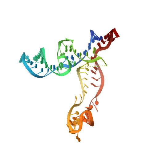
Crystal structure of Escherichia coli thiamine pyrophosphate-sensing riboswitch in the apo state.

Lee, H.K.Lee, Y.T.Fan, L.Wilt, H.M.Conrad, C.E.Yu, P.Zhang, J.Shi, G.Ji, X.Wang, Y.X.Stagno, J.R.

(2023) Structure 31: 848-859.e3

  • DOI: https://doi.org/10.1016/j.str.2023.05.003
  • Primary Citation of Related Structures:  
    8F4O

  • PubMed Abstract: 

    The thiamine pyrophosphate (TPP)-sensing riboswitch is one of the earliest discovered and most widespread riboswitches. Numerous structural studies have been reported for this riboswitch bound with various ligands. However, the ligand-free (apo) structure remains unknown. Here, we report a 3.1 Å resolution crystal structure of Escherichia coli TPP riboswitch in the apo state, which exhibits an extended, Y-shaped conformation further supported by small-angle X-ray scattering data and driven molecular dynamics simulations. The loss of ligand interactions results in helical uncoiling of P5 and disruption of the key tertiary interaction between the sensory domains. Opening of the aptamer propagates to the gene-regulatory P1 helix and generates the key conformational flexibility needed for the switching behavior. Much of the ligand-binding site at the three-way junction is unaltered, thereby maintaining a partially preformed pocket. Together, these results paint a dynamic picture of the ligand-induced conformational changes in TPP riboswitches that confer conditional gene regulation.


  • Organizational Affiliation

    Protein-Nucleic Acid Interaction Section, Center for Structural Biology, Center for Cancer Research, National Cancer Institute, Frederick, MD 21702, USA.


Macromolecules
Find similar nucleic acids by:  (by identity cutoff)  |  3D Structure
Entity ID: 1
MoleculeChains LengthOrganismImage
TPP riboswitch aptamer domain
A, B
83Escherichia coli
Sequence Annotations
Expand
  • Reference Sequence
Small Molecules
Ligands 3 Unique
IDChains Name / Formula / InChI Key2D Diagram3D Interactions
IRI
Query on IRI

Download Ideal Coordinates CCD File 
AA [auth B]
BA [auth B]
C [auth A]
CA [auth B]
D [auth A]
AA [auth B],
BA [auth B],
C [auth A],
CA [auth B],
D [auth A],
DA [auth B],
E [auth A],
EA [auth B],
F [auth A],
G [auth A],
H [auth A],
I [auth A],
J [auth A],
K [auth A],
L [auth A],
M [auth A],
N [auth A],
O [auth A],
P [auth A],
Q [auth A],
R [auth A],
S [auth A],
U [auth B],
V [auth B],
W [auth B],
X [auth B],
Y [auth B],
Z [auth B]
IRIDIUM HEXAMMINE ION
H18 Ir N6
CGMAOQLDNKCXGK-UHFFFAOYSA-N
PG4
Query on PG4

Download Ideal Coordinates CCD File 
GA [auth B]TETRAETHYLENE GLYCOL
C8 H18 O5
UWHCKJMYHZGTIT-UHFFFAOYSA-N
PGE
Query on PGE

Download Ideal Coordinates CCD File 
FA [auth B],
T [auth A]
TRIETHYLENE GLYCOL
C6 H14 O4
ZIBGPFATKBEMQZ-UHFFFAOYSA-N
Experimental Data & Validation

Experimental Data

  • Method: X-RAY DIFFRACTION
  • Resolution: 3.10 Å
  • R-Value Free: 0.317 
  • R-Value Work: 0.282 
  • R-Value Observed: 0.284 
  • Space Group: P 65 2 2
Unit Cell:
Length ( Å )Angle ( ˚ )
a = 139.264α = 90
b = 139.264β = 90
c = 97.905γ = 120
Software Package:
Software NamePurpose
PHENIXrefinement
Aimlessdata scaling
XDSdata reduction
PHASERphasing

Structure Validation

View Full Validation Report


Entry History & Funding Information

Deposition Data


Funding OrganizationLocationGrant Number
National Institutes of Health/National Cancer Institute (NIH/NCI)United States--

Revision History  (Full details and data files)

  • Version 1.0: 2023-05-17
    Type: Initial release
  • Version 1.1: 2023-06-21
    Changes: Database references
  • Version 1.2: 2023-07-19
    Changes: Database references
  • Version 1.3: 2023-10-25
    Changes: Data collection