Discovery of Potent Small-Molecule Inhibitors of WDR5-MYC Interaction.
Ding, J., Li, G., Liu, H., Liu, L., Lin, Y., Gao, J., Zhou, G., Shen, L., Zhao, M., Yu, Y., Guo, W., Hommel, U., Ottl, J., Blank, J., Aubin, N., Wei, Y., He, H., Sage, D.R., Atadja, P.W., Li, E., Jain, R.K., Tallarico, J.A., Canham, S.M., Chiang, Y.L., Wang, H.(2023) ACS Chem Biol 18: 34-40
- PubMed: 36594833 Search on PubMed
- DOI: https://doi.org/10.1021/acschembio.2c00843
- Primary Citation Related Structures:
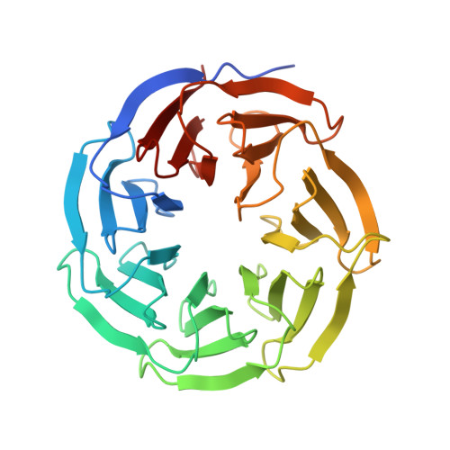
8F1G - PubMed Abstract:
WD repeat domain 5 (WDR5) is a member of the WD40-repeat protein family that plays a critical role in multiple processes. It is also a prominent target for pharmacological inhibition in diseases such as cancer, aging, and neurodegenerative disorders. Interactions between WDR5 and various partners are essential for sustaining its function. Most drug discovery efforts center on the WIN (WDR5 interaction motif) site of WDR5 that is responsible for the recruitment of WDR5 to chromatin. Here, we describe the discovery of novel WDR5 inhibitors for the other WBM (WDR5 binding motif) pocket on this scaffold protein, to disrupt WDR5 interaction with its binding partner MYC by high-throughput biochemical screening, subsequent molecule optimization, and biological assessment. These new WDR5 inhibitors provide useful probes for future investigations of WDR5 and an avenue for targeting WDR5 as a therapeutic strategy.
- Novartis Institutes for BioMedical Research, Cambridge, Massachusetts02139, United States.
Organizational Affiliation: