8F12 | pdb_00008f12

Structure of the MDM2 P53 binding domain in complex with H103, an all-D Helicon Polypeptide


Experimental Data Snapshot

  • Method: X-RAY DIFFRACTION
  • Resolution: 1.86 Å
  • R-Value Free: 
    0.241 (Depositor), 0.247 (DCC) 
  • R-Value Work: 
    0.215 (Depositor), 0.223 (DCC) 
  • R-Value Observed: 
    0.216 (Depositor) 

Starting Model: experimental
View more details

wwPDB Validation 3D Report Full Report

Validation slider image for 8F12

Ligand Structure Quality Assessment 


This is version 1.2 of the entry. See complete history

Literature

Macromolecule Content 

  • Total Structure Weight: 13.5 kDa 
  • Atom Count: 1,073 
  • Modeled Residue Count: 111 
  • Deposited Residue Count: 114 
  • Unique protein chains: 2

Macromolecules

Find similar proteins by:|  3D Structure
Entity ID: 1
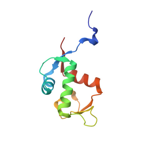
MoleculeChains  Sequence LengthOrganismDetailsImage
E3 ubiquitin-protein ligase Mdm295Homo sapiensMutation(s): 0 
Gene Names: MDM2
EC: 2.3.2.27
UniProt & NIH Common Fund Data Resources
Find proteins for Q00987 (Homo sapiens)
Explore Q00987 
Go to UniProtKB:  Q00987
PHAROS:  Q00987
GTEx:  ENSG00000135679 
Entity Groups
Sequence Clusters30% Identity50% Identity70% Identity90% Identity95% Identity100% Identity
UniProt GroupQ00987
Sequence Annotations
Expand
Reference Sequence
Find similar proteins by:  Sequence   |   3D Structure  
Entity ID: 2
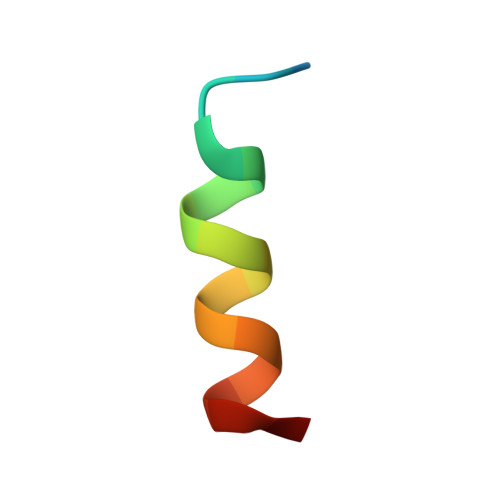
MoleculeChains  Sequence LengthOrganismDetailsImage
H10319synthetic constructMutation(s): 0 
Sequence Annotations
Expand
Reference Sequence

Small Molecules

Modified Residues  10 Unique
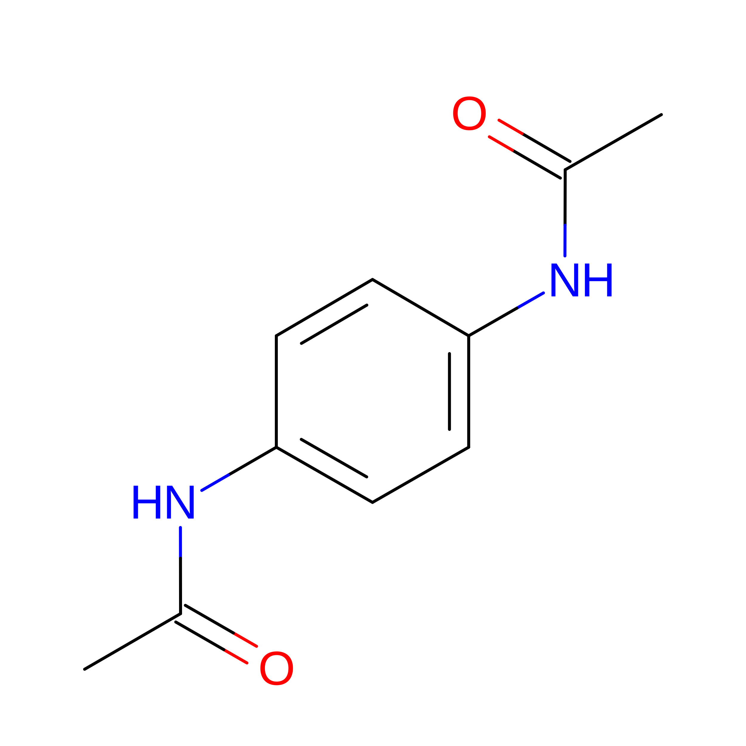
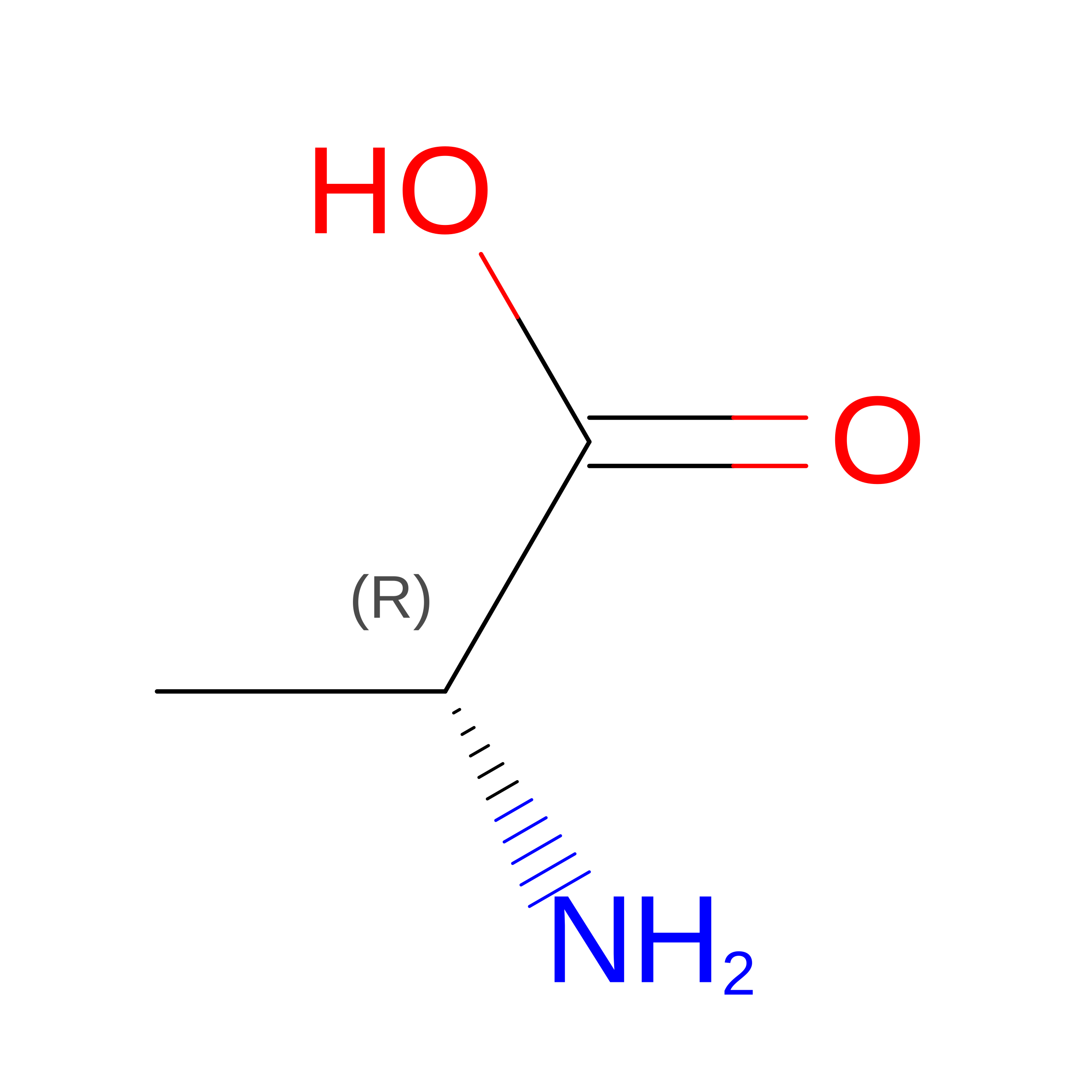
IDChains TypeFormula2D DiagramParent
DAL
Query on DAL
B
D-PEPTIDE LINKINGC3 H7 N O2

--

DAS
Query on DAS
B
D-PEPTIDE LINKINGC4 H7 N O4

--

DCY
Query on DCY
B
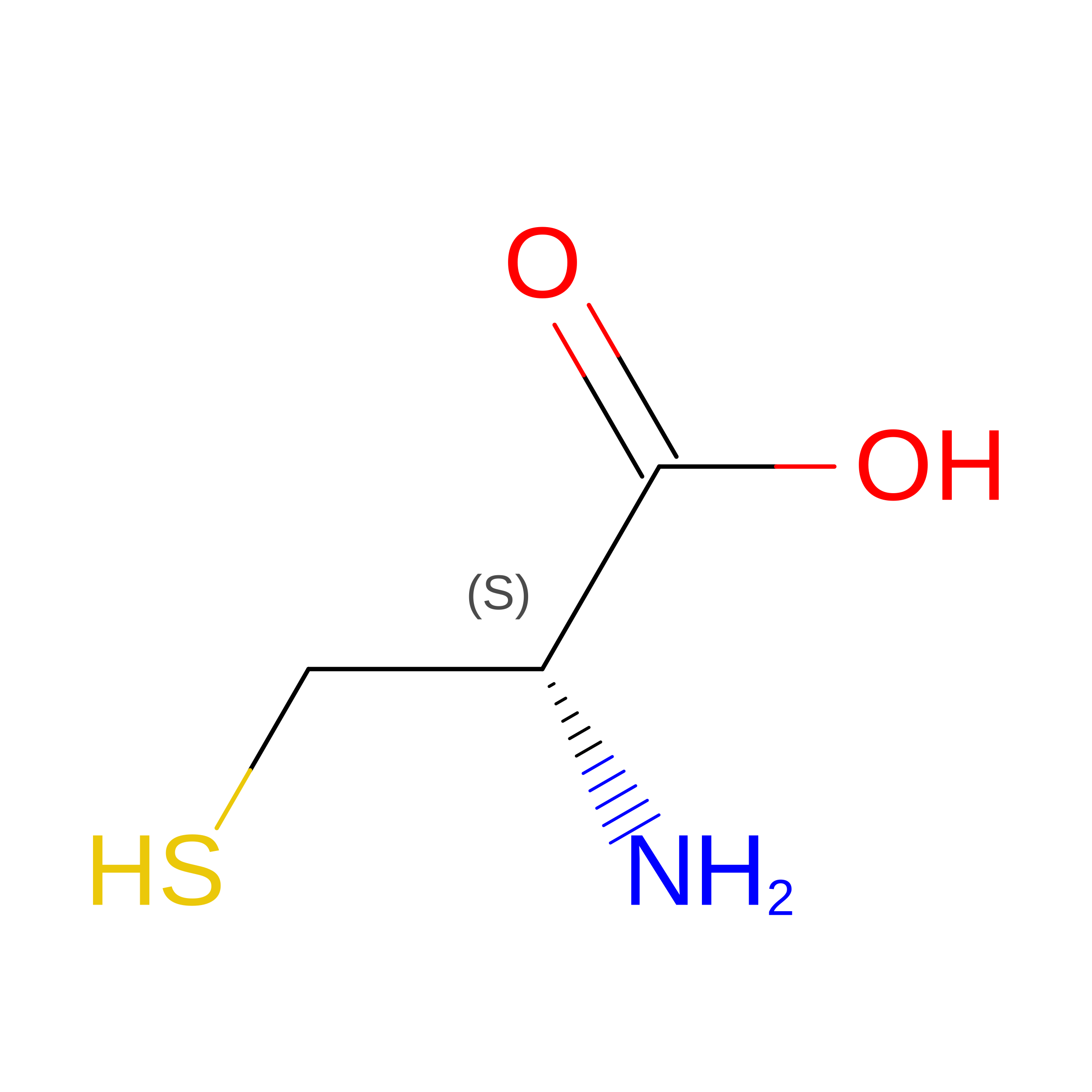
D-PEPTIDE LINKINGC3 H7 N O2 S

--

DGL
Query on DGL
B
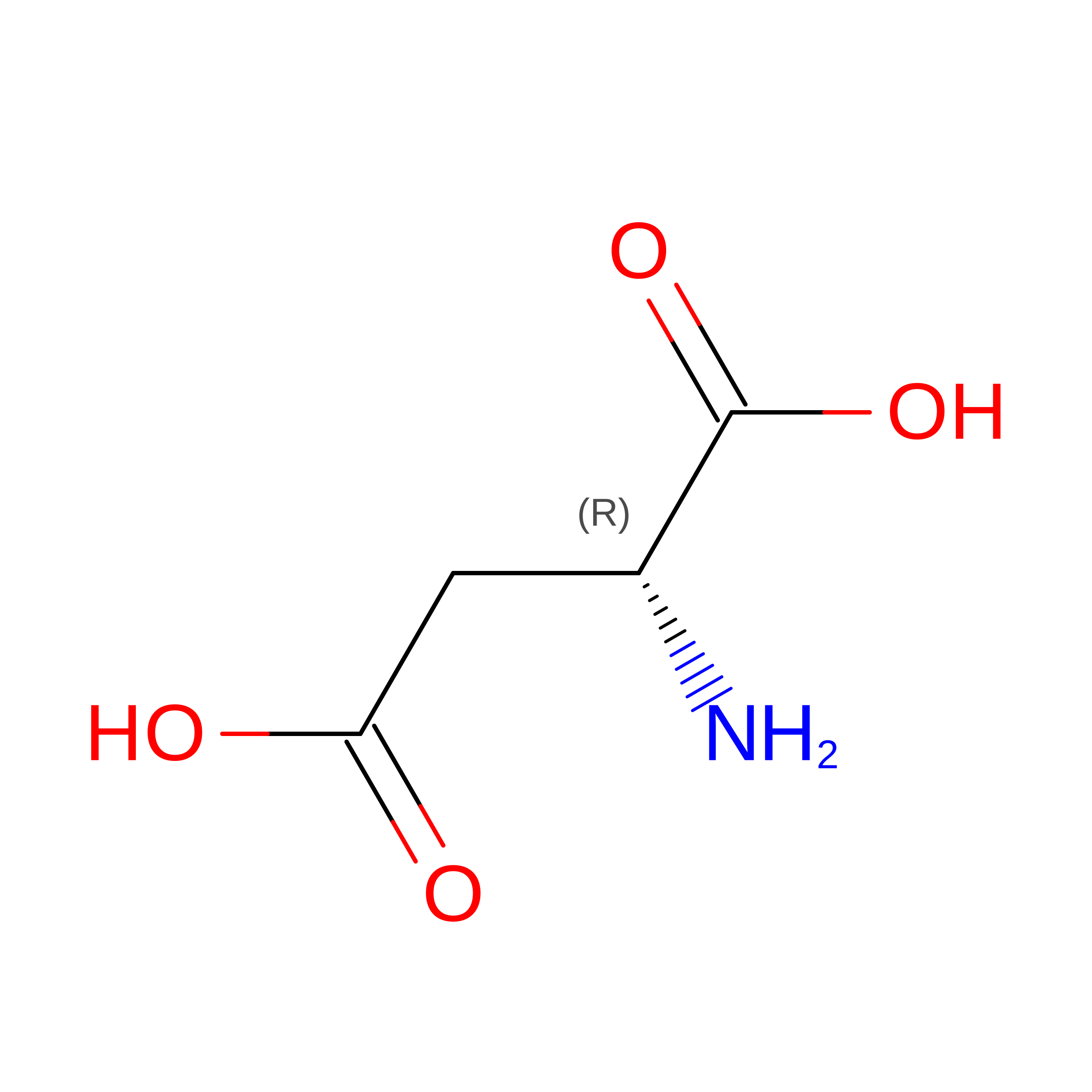
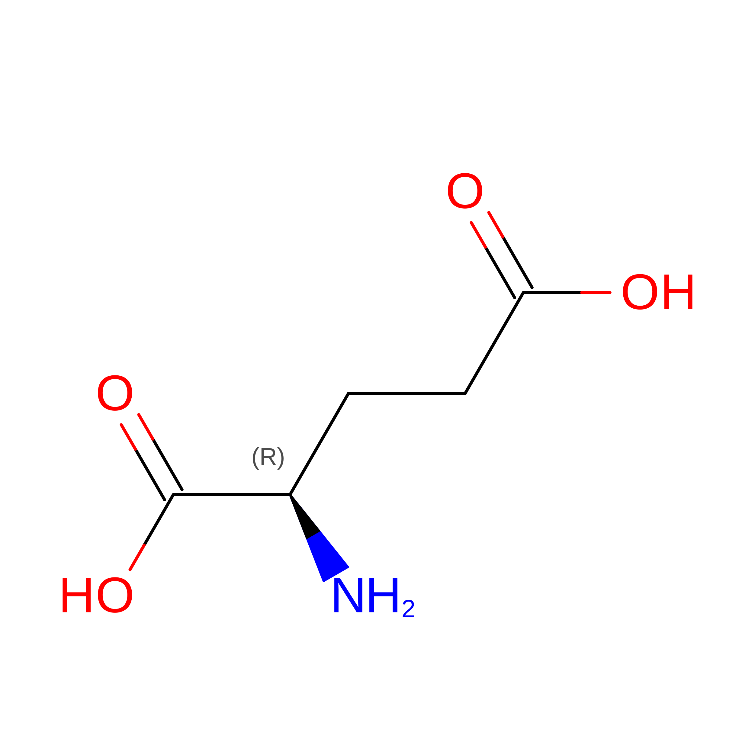
D-PEPTIDE LINKINGC5 H9 N O4

--

DGN
Query on DGN
B
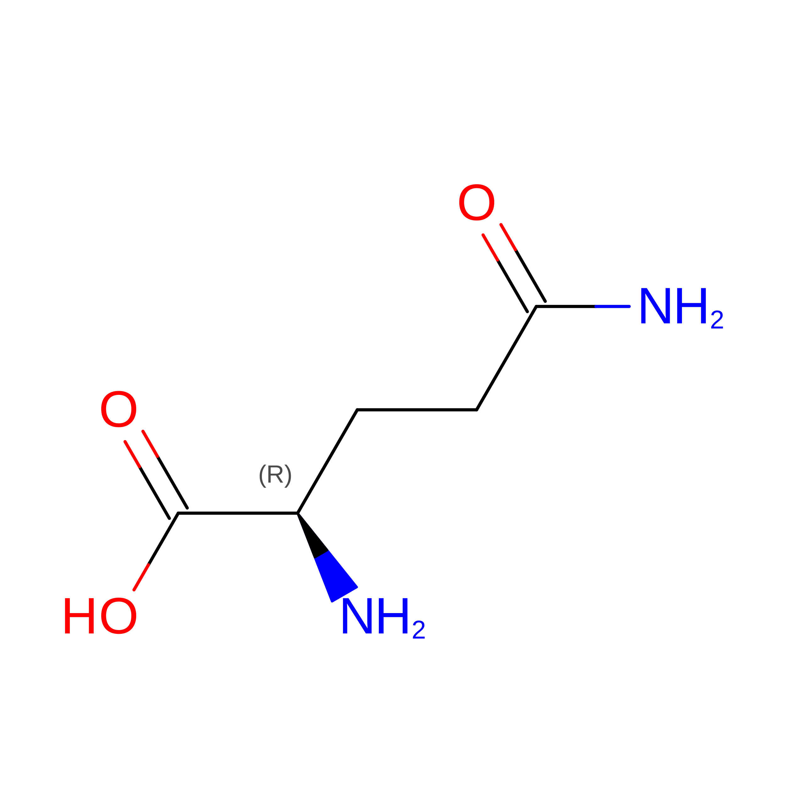
D-PEPTIDE LINKINGC5 H10 N2 O3

--

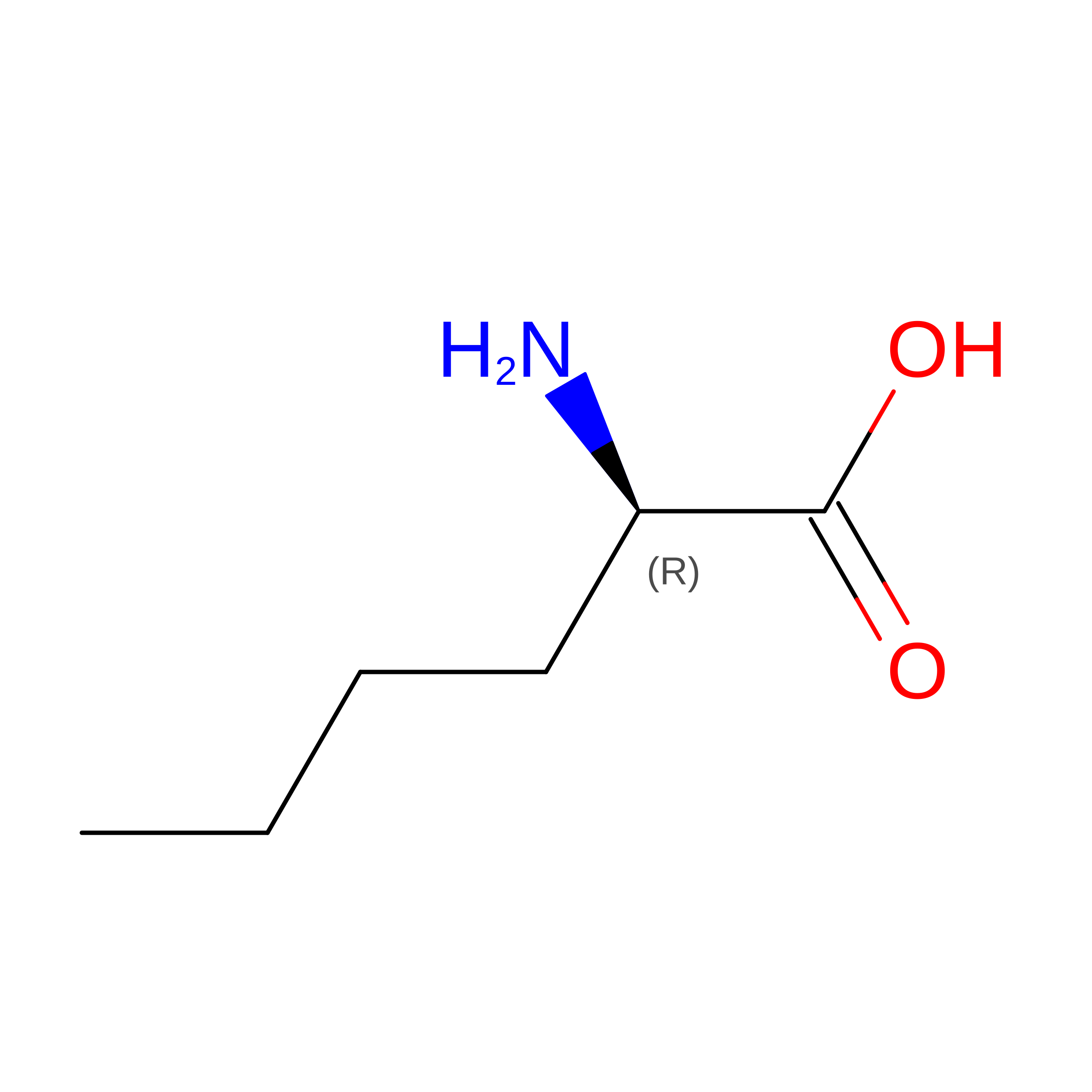
DNE
Query on DNE
B
D-PEPTIDE LINKINGC6 H13 N O2

--

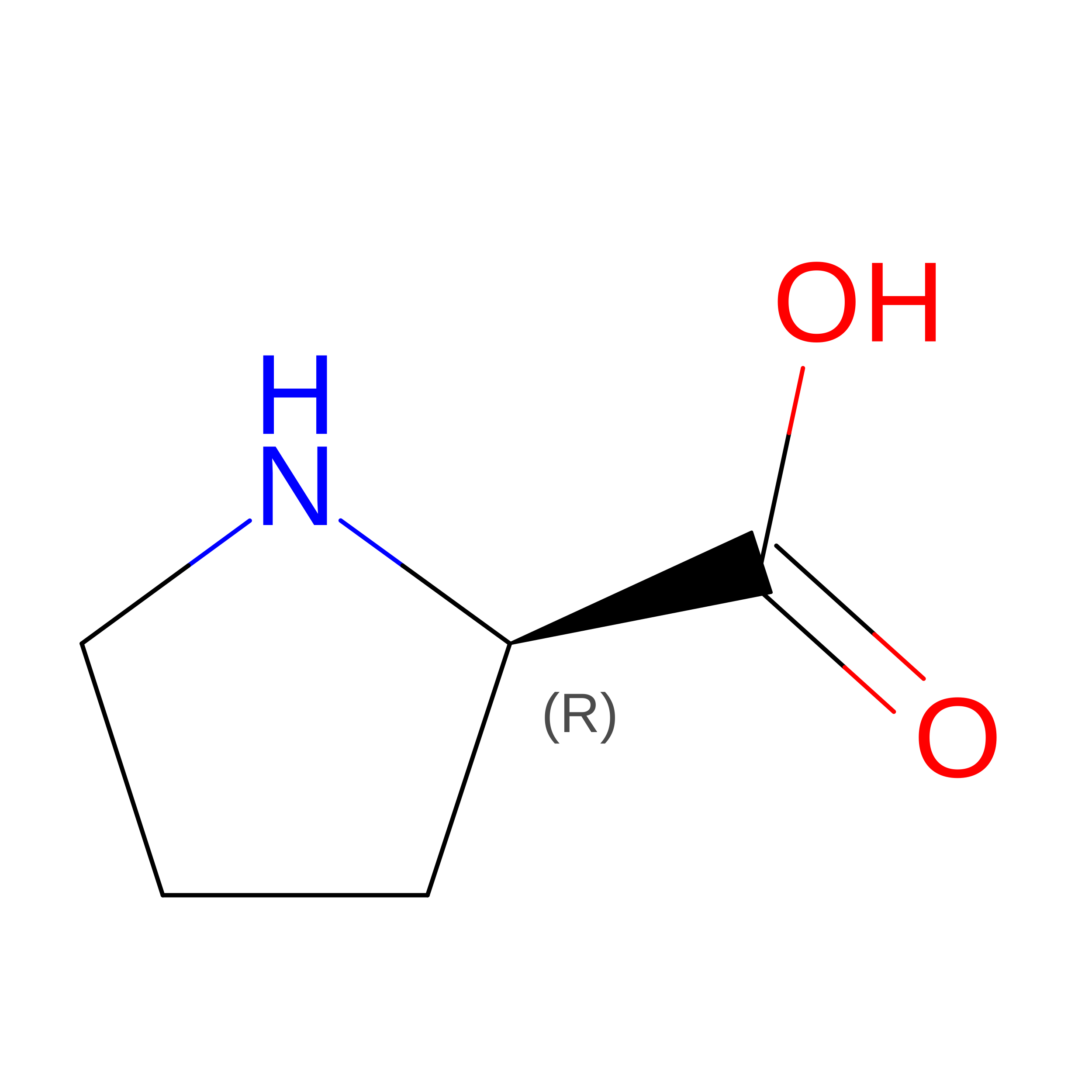
DPR
Query on DPR
B
D-PEPTIDE LINKINGC5 H9 N O2

--

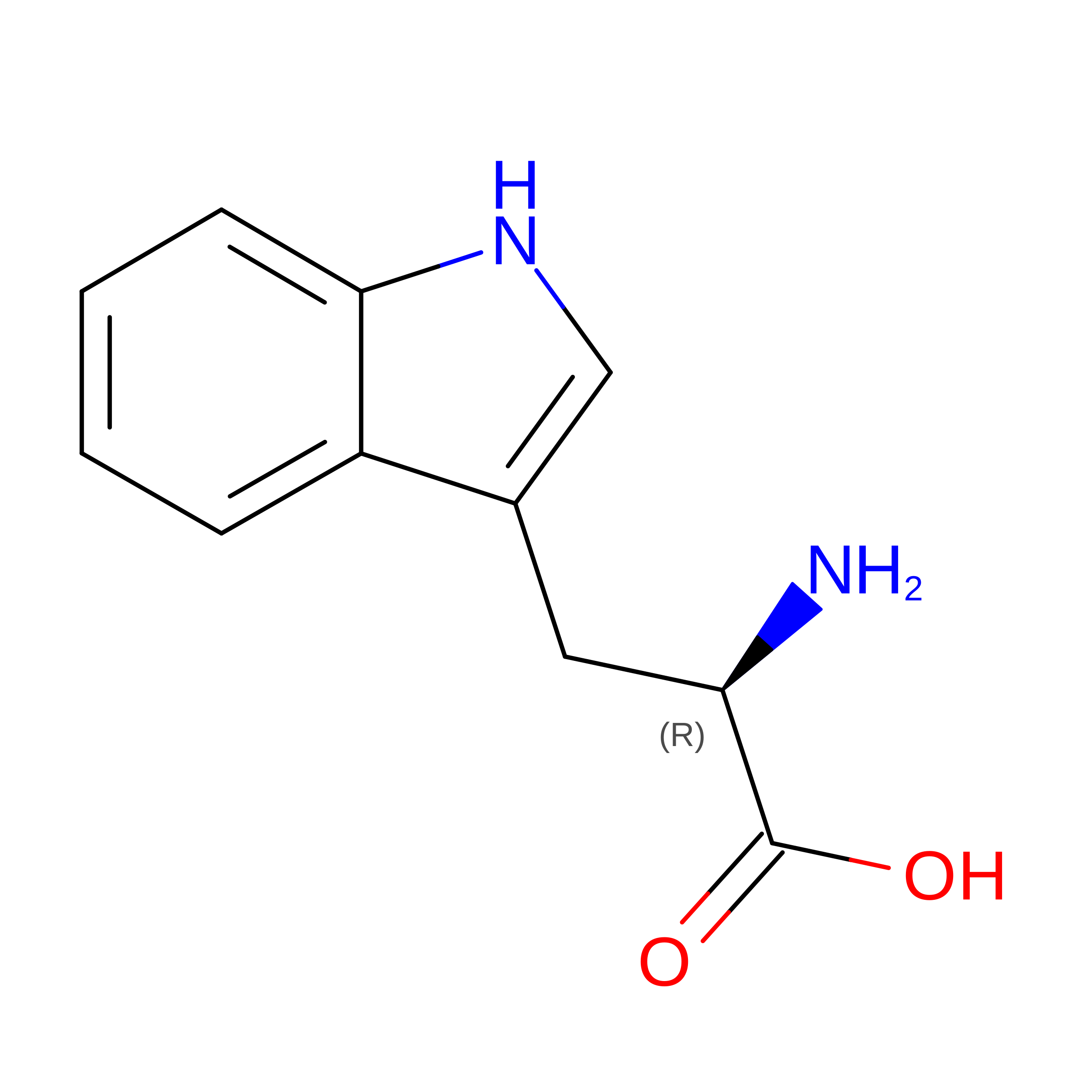
DTR
Query on DTR
B
D-PEPTIDE LINKINGC11 H12 N2 O2

--

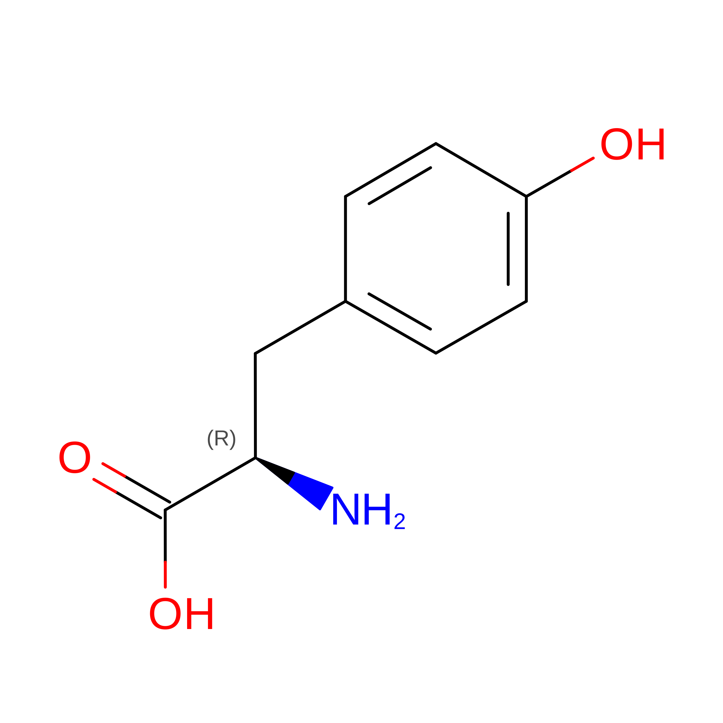
DTY
Query on DTY
B
D-PEPTIDE LINKINGC9 H11 N O3

--

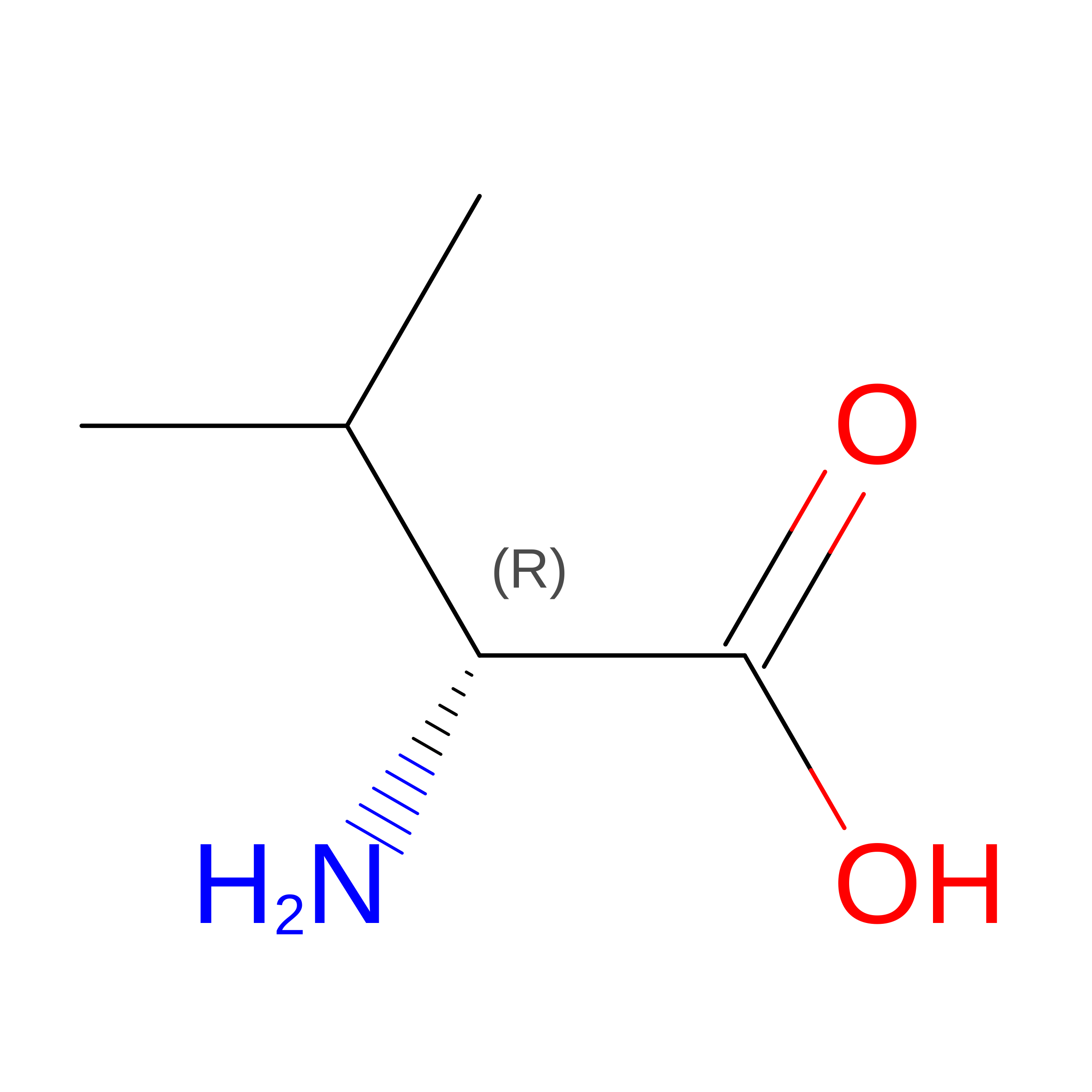
DVA
Query on DVA
B
D-PEPTIDE LINKINGC5 H11 N O2

--

Experimental Data & Validation

Experimental Data

  • Method: X-RAY DIFFRACTION
  • Resolution: 1.86 Å
  • R-Value Free:  0.241 (Depositor), 0.247 (DCC) 
  • R-Value Work:  0.215 (Depositor), 0.223 (DCC) 
  • R-Value Observed: 0.216 (Depositor) 
Space Group: P 43 21 2
Unit Cell:
Length ( Å )Angle ( ˚ )
a = 60.31α = 90
b = 60.31β = 90
c = 84.59γ = 90
Software Package:
Software NamePurpose
PHENIXrefinement
PDB_EXTRACTdata extraction
XDSdata reduction
Aimlessdata scaling
PHASERphasing

Structure Validation

View Full Validation Report


Ligand Structure Quality Assessment 


Entry History 

& Funding Information

Deposition Data


Funding OrganizationLocationGrant Number
Other privateUnited StatesFOG Pharmaceuticals
Other privateUnited StatesMIT School of Science Fellowship in Cancer Research
German Research Foundation (DFG)GermanyLE 4224/1-1

Revision History  (Full details and data files)

  • Version 1.0: 2023-02-15
    Type: Initial release
  • Version 1.1: 2023-10-25
    Changes: Data collection, Refinement description
  • Version 1.2: 2023-11-15
    Changes: Data collection