Structures of brain-derived 42-residue amyloid-beta fibril polymorphs with unusual molecular conformations and intermolecular interactions.
Lee, M., Yau, W.M., Louis, J.M., Tycko, R.(2023) Proc Natl Acad Sci U S A 120: e2218831120-e2218831120
- PubMed: 36893281 Search on PubMedSearch on PubMed Central
- DOI: https://doi.org/10.1073/pnas.2218831120
- Primary Citation Related Structures:
8EZD, 8EZE - PubMed Abstract:
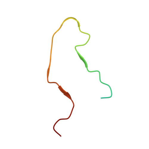
Fibrils formed by the 42-residue amyloid-β peptide (Aβ42), a main component of amyloid deposits in Alzheimer's disease (AD), are known to be polymorphic, i.e., to contain multiple possible molecular structures. Previous studies of Aβ42 fibrils, including fibrils prepared entirely in vitro or extracted from brain tissue and using solid-state NMR (ssNMR) or cryogenic electron microscopy (cryo-EM) methods, have found polymorphs with differences in amino acid sidechain orientations, lengths of structurally ordered segments, and contacts between cross-β subunit pairs within a single filament. Despite these differences, Aβ42 molecules adopt a common S-shaped conformation in all previously described high-resolution Aβ42 fibril structures. Here we report two cryo-EM-based structures of Aβ42 fibrils that are qualitatively different, in samples derived from AD brain tissue by seeded growth. In type A fibrils, residues 12 to 42 adopt a ν-shaped conformation, with both intra-subunit and intersubunit hydrophobic contacts to form a compact core. In type B fibrils, residues 2 to 42 adopt an υ-shaped conformation, with only intersubunit contacts and internal pores. Type A and type B fibrils have opposite helical handedness. Cryo-EM density maps and molecular dynamics simulations indicate intersubunit K16-A42 salt bridges in type B fibrils and partially occupied K28-A42 salt bridges in type A fibrils. The coexistence of two predominant polymorphs, with differences in N-terminal dynamics, is supported by ssNMR data, as is faithful propagation of structures from first-generation to second-generation brain-seeded Aβ42 fibril samples. These results demonstrate that Aβ42 fibrils can exhibit a greater range of structural variations than seen in previous studies.
- Laboratory of Chemical Physics, National Institute of Diabetes and Digestive and Kidney Diseases, NIH, Bethesda, MD 20892-0520.
Organizational Affiliation: