8EVZ

DdlB from Pseudomonas aeruginosa PAO1 in complex with ADP and phosphorylated D-cycloserine


Experimental Data Snapshot

  • Method: X-RAY DIFFRACTION
  • Resolution: 2.45 Å
  • R-Value Free: 0.245 
  • R-Value Work: 0.204 
  • R-Value Observed: 0.206 

wwPDB Validation   3D Report Full Report


Ligand Structure Quality Assessment 


This is version 1.2 of the entry. See complete history


Literature

Comparative functional and structural analysis of Pseudomonas aeruginosa d-alanine-d-alanine ligase isoforms as prospective antibiotic targets.

Pederick, J.L.Woolman, J.C.Bruning, J.B.

(2023) FEBS J 290: 5536-5553

  • DOI: https://doi.org/10.1111/febs.16932
  • Primary Citation of Related Structures:  
    8EVV, 8EVW, 8EVX, 8EVY, 8EVZ

  • PubMed Abstract: 

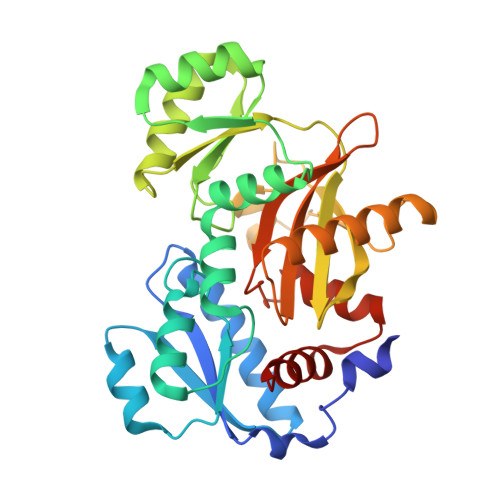
    Pseudomonas aeruginosa is a major human pathogen in the healthcare setting. The emergence of multi-drug-resistant and extensive drug-resistant P. aeruginosa is of great concern, and clearly indicates that new alternatives to current first-line antibiotics are required in the future. Inhibition of d-alanine-d-alanine production presents as a promising avenue as it is a key component in the essential process of cell wall biosynthesis. In P. aeruginosa, d-alanine-d-alanine production is facilitated by two isoforms, d-alanine-d-alanine ligase A (PaDdlA) and d-alanine-d-alanine ligase B (PaDdlA), but neither enzyme has been individually characterised to date. Here, we present the functional and structural characterisation of PaDdlA and PaDdlB, and assess their potential as antibiotic targets. This was achieved using a combination of in vitro enzyme-activity assays and X-ray crystallography. The former revealed that both isoforms effectively catalyse d-alanine-d-alanine production with near identical efficiency, and that this is effectively disrupted by the model d-alanine-d-alanine ligase inhibitor, d-cycloserine. Next, each isoform was co-crystallised with ATP and either d-alanine-d-alanine or d-cycloserine, allowing direct comparison of the key structural features. Both isoforms possess the same structural architecture and share a high level of conservation within the active site. Although residues forming the d-alanine pocket are completely conserved, the ATP-binding pocket possesses several amino acid substitutions resulting in a differing chemical environment around the ATP adenine base. Together, these findings support that the discovery of dual PaDdlA/PaDdlB competitive inhibitors is a viable approach for developing new antibiotics against P. aeruginosa.


  • Organizational Affiliation

    Institute for Photonics and Advanced Sensing (IPAS), School of Biological Sciences, The University of Adelaide, SA, Australia.


Macromolecules
Find similar proteins by:  (by identity cutoff)  |  3D Structure
Entity ID: 1
MoleculeChains Sequence LengthOrganismDetailsImage
D-alanine--D-alanine ligase B
A, B, C
320Pseudomonas aeruginosa PAO1Mutation(s): 0 
Gene Names: ddlBPA4410
EC: 6.3.2.4
UniProt
Find proteins for Q9LCT6 (Pseudomonas aeruginosa (strain ATCC 15692 / DSM 22644 / CIP 104116 / JCM 14847 / LMG 12228 / 1C / PRS 101 / PAO1))
Explore Q9LCT6 
Go to UniProtKB:  Q9LCT6
Entity Groups  
Sequence Clusters30% Identity50% Identity70% Identity90% Identity95% Identity100% Identity
UniProt GroupQ9LCT6
Sequence Annotations
Expand
  • Reference Sequence
Small Molecules
Ligands 3 Unique
IDChains Name / Formula / InChI Key2D Diagram3D Interactions
ADP (Subject of Investigation/LOI)
Query on ADP

Download Ideal Coordinates CCD File 
F [auth A],
J [auth B],
N [auth C]
ADENOSINE-5'-DIPHOSPHATE
C10 H15 N5 O10 P2
XTWYTFMLZFPYCI-KQYNXXCUSA-N
DS0 (Subject of Investigation/LOI)
Query on DS0

Download Ideal Coordinates CCD File 
G [auth A],
K [auth B],
O [auth C]
[(4R)-4-azanyl-4,5-dihydro-1,2-oxazol-3-yl] dihydrogen phosphate
C3 H7 N2 O5 P
SFMNESRSKJTEMJ-UWTATZPHSA-N
MG
Query on MG

Download Ideal Coordinates CCD File 
D [auth A]
E [auth A]
H [auth B]
I [auth B]
L [auth C]
D [auth A],
E [auth A],
H [auth B],
I [auth B],
L [auth C],
M [auth C]
MAGNESIUM ION
Mg
JLVVSXFLKOJNIY-UHFFFAOYSA-N
Experimental Data & Validation

Experimental Data

  • Method: X-RAY DIFFRACTION
  • Resolution: 2.45 Å
  • R-Value Free: 0.245 
  • R-Value Work: 0.204 
  • R-Value Observed: 0.206 
  • Space Group: C 2 2 21
Unit Cell:
Length ( Å )Angle ( ˚ )
a = 90.664α = 90
b = 199.046β = 90
c = 177.084γ = 90
Software Package:
Software NamePurpose
PHENIXrefinement
XDSdata reduction
Aimlessdata scaling
PDB_EXTRACTdata extraction
PHASERphasing

Structure Validation

View Full Validation Report


Ligand Structure Quality Assessment 


Entry History & Funding Information

Deposition Data


Funding OrganizationLocationGrant Number
Not funded--

Revision History  (Full details and data files)

  • Version 1.0: 2023-08-30
    Type: Initial release
  • Version 1.1: 2023-09-06
    Changes: Database references
  • Version 1.2: 2023-12-20
    Changes: Database references