8EUW | pdb_00008euw

Cryo-EM structure of HIV-1 BG505 DS-SOSIP ENV trimer bound to VRC34.01-MM28 FAB


Experimental Data Snapshot

  • Method: ELECTRON MICROSCOPY
  • Resolution: 2.70 Å
  • Aggregation State: PARTICLE 
  • Reconstruction Method: SINGLE PARTICLE 

wwPDB Validation 3D Report Full Report

Validation slider image for 8EUW

This is version 1.3 of the entry. See complete history

Literature

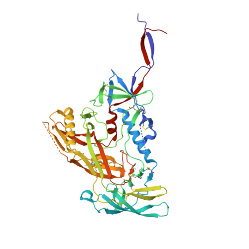
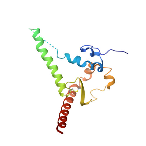
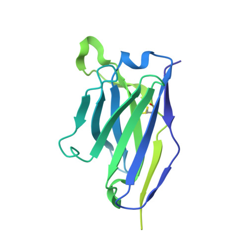
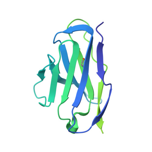
Antibody-directed evolution reveals a mechanism for enhanced neutralization at the HIV-1 fusion peptide site.

Banach, B.B.Pletnev, S.Olia, A.S.Xu, K.Zhang, B.Rawi, R.Bylund, T.Doria-Rose, N.A.Nguyen, T.D.Fahad, A.S.Lee, M.Lin, B.C.Liu, T.Louder, M.K.Madan, B.McKee, K.O'Dell, S.Sastry, M.Schon, A.Bui, N.Shen, C.H.Wolfe, J.R.Chuang, G.Y.Mascola, J.R.Kwong, P.D.DeKosky, B.J.

(2023) Nat Commun 14: 7593-7593

  • DOI: https://doi.org/10.1038/s41467-023-42098-5
  • Primary Citation Related Structures: 
    8ELI, 8EUU, 8EUV, 8EUW, 8F7Z

  • PubMed Abstract: 

    The HIV-1 fusion peptide (FP) represents a promising vaccine target, but global FP sequence diversity among circulating strains has limited anti-FP antibodies to ~60% neutralization breadth. Here we evolve the FP-targeting antibody VRC34.01 in vitro to enhance FP-neutralization using site saturation mutagenesis and yeast display. Successive rounds of directed evolution by iterative selection of antibodies for binding to resistant HIV-1 strains establish a variant, VRC34.01_mm28, as a best-in-class antibody with 10-fold enhanced potency compared to the template antibody and ~80% breadth on a cross-clade 208-strain neutralization panel. Structural analyses demonstrate that the improved paratope expands the FP binding groove to accommodate diverse FP sequences of different lengths while also recognizing the HIV-1 Env backbone. These data reveal critical antibody features for enhanced neutralization breadth and potency against the FP site of vulnerability and accelerate clinical development of broad HIV-1 FP-targeting vaccines and therapeutics.


  • Organizational Affiliation
    • Bioengineering Graduate Program, The University of Kansas, Lawrence, KS, 66045, USA.

Macromolecule Content 

  • Total Structure Weight: 371.5 kDa 
  • Atom Count: 20,040 
  • Modeled Residue Count: 2,400 
  • Deposited Residue Count: 3,207 
  • Unique protein chains: 4

Macromolecules

Find similar proteins by:|  3D Structure
Entity ID: 1
MoleculeChains  Sequence LengthOrganismDetailsImage
Envelope glycoprotein gp120
A, C, E
481Human immunodeficiency virus 1Mutation(s): 0 
Gene Names: env
UniProt
Find proteins for Q2N0S6 (Human immunodeficiency virus type 1)
Explore Q2N0S6 
Go to UniProtKB:  Q2N0S6
Entity Groups
Sequence Clusters30% Identity50% Identity70% Identity90% Identity95% Identity100% Identity
UniProt GroupQ2N0S6
Glycosylation
Glycosylation Sites: 15
Sequence Annotations
Expand
Reference Sequence
Find similar proteins by:|  3D Structure
Entity ID: 2
MoleculeChains  Sequence LengthOrganismDetailsImage
Envelope glycoprotein gp41
B, D, F
153Human immunodeficiency virus 1Mutation(s): 0 
Gene Names: env
UniProt
Find proteins for Q2N0S6 (Human immunodeficiency virus type 1)
Explore Q2N0S6 
Go to UniProtKB:  Q2N0S6
Entity Groups
Sequence Clusters30% Identity50% Identity70% Identity90% Identity95% Identity100% Identity
UniProt GroupQ2N0S6
Sequence Annotations
Expand
Reference Sequence
Find similar proteins by:|  3D Structure
Entity ID: 3
MoleculeChains  Sequence LengthOrganismDetailsImage
VRC34.01-MM28 FAB variable heavy chain
G, I, K
223Homo sapiensMutation(s): 0 
Find similar proteins by:|  3D Structure
Entity ID: 4
MoleculeChains  Sequence LengthOrganismDetailsImage
VRC34.01-MM28 FAB variable light chain
H, J, L
212Homo sapiensMutation(s): 0 

Oligosaccharides

Help  
Entity ID: 6
MoleculeChains Length2D Diagram GlycosylationD Interactions
beta-D-mannopyranose-(1-4)-2-acetamido-2-deoxy-beta-D-glucopyranose-(1-4)-2-acetamido-2-deoxy-beta-D-glucopyranoseEA [auth d],
O [auth N],
W [auth U]
3N-Glycosylation
Glycosylation Resources
GlyTouCan: G15407YE
GlyCosmos: G15407YE
GlyGen: G15407YE
Entity ID: 7
MoleculeChains Length2D Diagram GlycosylationD Interactions
alpha-D-mannopyranose-(1-2)-alpha-D-mannopyranose-(1-3)-beta-D-mannopyranose-(1-4)-2-acetamido-2-deoxy-beta-D-glucopyranose-(1-4)-2-acetamido-2-deoxy-beta-D-glucopyranoseGA [auth g],
Q [auth P],
Y [auth W]
5N-Glycosylation
Glycosylation Resources
GlyTouCan: G42227JK
GlyCosmos: G42227JK
GlyGen: G42227JK

Small Molecules

Ligands 1 Unique
IDChains Name / Formula / InChI Key2D Diagram3D Interactions
NAG

Query on NAG


Download:Ideal Coordinates CCD File
AB [auth E]
BB [auth E]
CB [auth E]
DB [auth E]
EB [auth E]
AB [auth E],
BB [auth E],
CB [auth E],
DB [auth E],
EB [auth E],
KA [auth A],
LA [auth A],
MA [auth A],
NA [auth A],
OA [auth A],
PA [auth A],
QA [auth A],
RA [auth C],
SA [auth C],
TA [auth C],
UA [auth C],
VA [auth C],
WA [auth C],
XA [auth C],
YA [auth E],
ZA [auth E]
2-acetamido-2-deoxy-beta-D-glucopyranose
C8 H15 N O6
OVRNDRQMDRJTHS-FMDGEEDCSA-N

Experimental Data & Validation

Experimental Data

  • Method: ELECTRON MICROSCOPY
  • Resolution: 2.70 Å
  • Aggregation State: PARTICLE 
  • Reconstruction Method: SINGLE PARTICLE 
EM Software:
TaskSoftware PackageVersion
MODEL REFINEMENTPHENIX1.20
RECONSTRUCTIONcryoSPARC3.3

Structure Validation

View Full Validation Report


Entry History 

& Funding Information

Deposition Data


Funding OrganizationLocationGrant Number
National Institutes of Health/National Institute Of Allergy and Infectious Diseases (NIH/NIAID)United States--

Revision History  (Full details and data files)

  • Version 1.0: 2023-09-27
    Type: Initial release
  • Version 1.1: 2024-02-14
    Changes: Database references
  • Version 1.2: 2024-11-13
    Changes: Data collection, Structure summary
  • Version 1.3: 2025-03-26
    Changes: Data collection