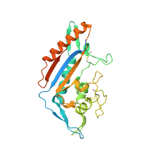
8EUS

Crystal structure of NPC1 luminal domain C


Experimental Data Snapshot

  • Method: X-RAY DIFFRACTION
  • Resolution: 2.30 Å
  • R-Value Free: 0.229 
  • R-Value Work: 0.191 
  • R-Value Observed: 0.193 

wwPDB Validation   3D Report Full Report


This is version 1.1 of the entry. See complete history


Literature

Purification and structure of luminal domain C of human Niemann-Pick C1 protein.

Odongo, L.Zadrozny, K.K.Diehl, W.E.Luban, J.White, J.M.Ganser-Pornillos, B.K.Tamm, L.K.Pornillos, O.

(2023) Acta Crystallogr F Struct Biol Commun 79: 45-50

  • DOI: https://doi.org/10.1107/S2053230X23000705
  • Primary Citation of Related Structures:  
    8EUS

  • PubMed Abstract: 

    Niemann-Pick C1 protein (NPC1) is a membrane protein that primarily resides in late endosomes and lysosomes, and plays an important role in cholesterol homeostasis in the cell. The second luminal domain of NPC1 (NPC1-C) serves as the intracellular receptor for Ebola and Marburg viruses. Here, the recombinant production of nonglycosylated and glycosylated NPC1-C and a new crystal form of the nonglycosylated protein are reported. The crystals belonged to space group P2 1 and diffracted to 2.3 Å resolution. The structure is similar to other reported structures of NPC1-C, with differences observed in the protruding loops when compared with NPC1-C in complex with Ebola virus glycoprotein or NPC2.


  • Organizational Affiliation

    Center for Membrane and Cell Physiology, University of Virginia, Charlottesville, VA 22908, USA.


Macromolecules
Find similar proteins by:  (by identity cutoff)  |  3D Structure
Entity ID: 1
MoleculeChains Sequence LengthOrganismDetailsImage
NPC intracellular cholesterol transporter 1A [auth C],
B [auth A]
262Homo sapiensMutation(s): 0 
Gene Names: NPC1
Membrane Entity: Yes 
UniProt & NIH Common Fund Data Resources
Find proteins for O15118 (Homo sapiens)
Explore O15118 
Go to UniProtKB:  O15118
PHAROS:  O15118
GTEx:  ENSG00000141458 
Entity Groups  
Sequence Clusters30% Identity50% Identity70% Identity90% Identity95% Identity100% Identity
UniProt GroupO15118
Sequence Annotations
Expand
  • Reference Sequence
Experimental Data & Validation

Experimental Data

  • Method: X-RAY DIFFRACTION
  • Resolution: 2.30 Å
  • R-Value Free: 0.229 
  • R-Value Work: 0.191 
  • R-Value Observed: 0.193 
  • Space Group: P 1 21 1
Unit Cell:
Length ( Å )Angle ( ˚ )
a = 53.145α = 90
b = 65.207β = 91.69
c = 68.534γ = 90
Software Package:
Software NamePurpose
PHENIXrefinement
PHENIXrefinement
HKL-2000data reduction
HKL-2000data scaling
REFMACphasing

Structure Validation

View Full Validation Report


Entry History & Funding Information

Deposition Data


Funding OrganizationLocationGrant Number
National Institutes of Health/National Institute Of Allergy and Infectious Diseases (NIH/NIAID)United StatesR01 AI30557

Revision History  (Full details and data files)

  • Version 1.0: 2023-02-22
    Type: Initial release
  • Version 1.1: 2023-10-25
    Changes: Data collection, Refinement description