8ERC | pdb_00008erc

Human Membrane-bound O-acyltransferase 7


Experimental Data Snapshot

  • Method: ELECTRON MICROSCOPY
  • Resolution: 3.70 Å
  • Aggregation State: 2D ARRAY 
  • Reconstruction Method: SINGLE PARTICLE 

wwPDB Validation 3D Report Full Report

Validation slider image for 8ERC

This is version 1.1 of the entry. See complete history

Literature

The structure of phosphatidylinositol remodeling MBOAT7 reveals its catalytic mechanism and enables inhibitor identification.

Wang, K.Lee, C.W.Sui, X.Kim, S.Wang, S.Higgs, A.B.Baublis, A.J.Voth, G.A.Liao, M.Walther, T.C.Farese Jr., R.V.

(2023) Nat Commun 14: 3533-3533

  • DOI: https://doi.org/10.1038/s41467-023-38932-5
  • Primary Citation Related Structures: 
    8ERC

  • PubMed Abstract: 

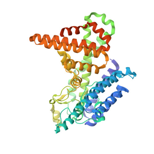
    Cells remodel glycerophospholipid acyl chains via the Lands cycle to adjust membrane properties. Membrane-bound O-acyltransferase (MBOAT) 7 acylates lyso-phosphatidylinositol (lyso-PI) with arachidonyl-CoA. MBOAT7 mutations cause brain developmental disorders, and reduced expression is linked to fatty liver disease. In contrast, increased MBOAT7 expression is linked to hepatocellular and renal cancers. The mechanistic basis of MBOAT7 catalysis and substrate selectivity are unknown. Here, we report the structure and a model for the catalytic mechanism of human MBOAT7. Arachidonyl-CoA and lyso-PI access the catalytic center through a twisted tunnel from the cytosol and lumenal sides, respectively. N-terminal residues on the ER lumenal side determine phospholipid headgroup selectivity: swapping them between MBOATs 1, 5, and 7 converts enzyme specificity for different lyso-phospholipids. Finally, the MBOAT7 structure and virtual screening enabled identification of small-molecule inhibitors that may serve as lead compounds for pharmacologic development.


  • Organizational Affiliation
    • Department of Molecular Metabolism, Harvard T.H. Chan School of Public Health, Boston, MA, USA.

Macromolecule Content 

  • Total Structure Weight: 54.2 kDa 
  • Atom Count: 3,510 
  • Modeled Residue Count: 441 
  • Deposited Residue Count: 484 
  • Unique protein chains: 1

Macromolecules

Find similar proteins by:|  3D Structure
Entity ID: 1
MoleculeChains  Sequence LengthOrganismDetailsImage
Lysophospholipid acyltransferase 7484Homo sapiensMutation(s): 0 
Gene Names: MBOAT7BB1LENG4OACT7
EC: 2.3.1
Membrane Entity: Yes 
UniProt & NIH Common Fund Data Resources
Find proteins for Q96N66 (Homo sapiens)
Explore Q96N66 
Go to UniProtKB:  Q96N66
PHAROS:  Q96N66
GTEx:  ENSG00000125505 
Entity Groups
Sequence Clusters30% Identity50% Identity70% Identity90% Identity95% Identity100% Identity
UniProt GroupQ96N66
Sequence Annotations
Expand
Reference Sequence

Experimental Data & Validation

Experimental Data

  • Method: ELECTRON MICROSCOPY
  • Resolution: 3.70 Å
  • Aggregation State: 2D ARRAY 
  • Reconstruction Method: SINGLE PARTICLE 
EM Software:
TaskSoftware PackageVersion
MODEL REFINEMENTPHENIX

Structure Validation

View Full Validation Report


Entry History 

& Funding Information

Deposition Data


Funding OrganizationLocationGrant Number
National Institutes of Health/National Center for Research Resources (NIH/NCRR)United StatesR01GM141050
Howard Hughes Medical Institute (HHMI)United States--

Revision History  (Full details and data files)

  • Version 1.0: 2023-09-27
    Type: Initial release
  • Version 1.1: 2025-06-04
    Changes: Data collection, Structure summary