8EPM

Human R-type voltage-gated calcium channel Cav2.3 CH2II-deleted mutant at 3.1 Angstrom resolution


Experimental Data Snapshot

  • Method: ELECTRON MICROSCOPY
  • Resolution: 3.10 Å
  • Aggregation State: PARTICLE 
  • Reconstruction Method: SINGLE PARTICLE 

wwPDB Validation   3D Report Full Report


This is version 1.1 of the entry. See complete history


Literature

Structures of the R-type human Ca v 2.3 channel reveal conformational crosstalk of the intracellular segments.

Yao, X.Wang, Y.Wang, Z.Fan, X.Wu, D.Huang, J.Mueller, A.Gao, S.Hu, M.Robinson, C.V.Yu, Y.Gao, S.Yan, N.

(2022) Nat Commun 13: 7358-7358

  • DOI: https://doi.org/10.1038/s41467-022-35026-6
  • Primary Citation of Related Structures:  
    8EPL, 8EPM

  • PubMed Abstract: 

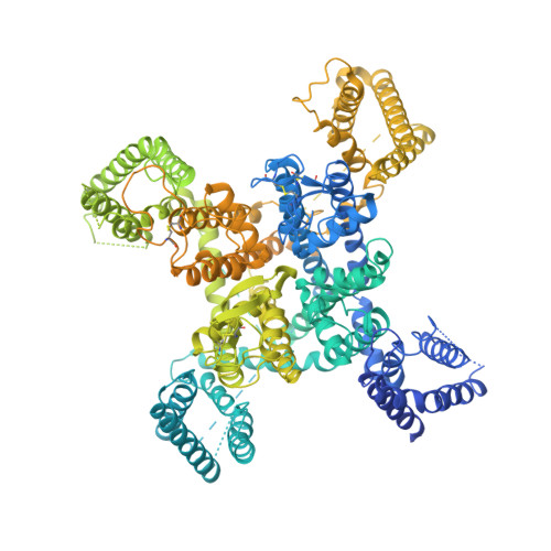
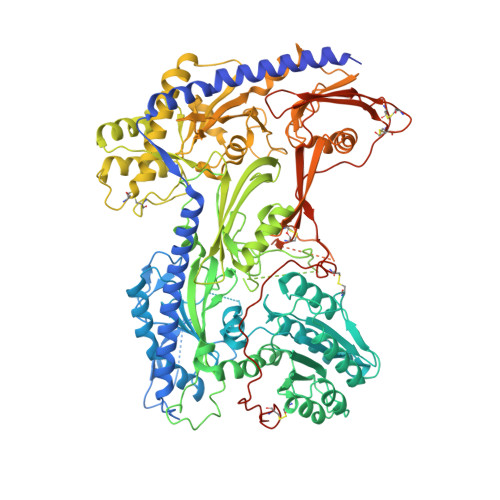
    The R-type voltage-gated Ca 2+ (Ca v ) channels Ca v 2.3, widely expressed in neuronal and neuroendocrine cells, represent potential drug targets for pain, seizures, epilepsy, and Parkinson's disease. Despite their physiological importance, there have lacked selective small-molecule inhibitors targeting these channels. High-resolution structures may aid rational drug design. Here, we report the cryo-EM structure of human Ca v 2.3 in complex with α2δ-1 and β3 subunits at an overall resolution of 3.1 Å. The structure is nearly identical to that of Ca v 2.2, with VSD II in the down state and the other three VSDs up. A phosphatidylinositol 4,5-bisphosphate (PIP2) molecule binds to the interface of VSD II and the tightly closed pore domain. We also determined the cryo-EM structure of a Ca v 2.3 mutant in which a Ca v 2-unique cytosolic helix in repeat II (designated the CH2 II helix) is deleted. This mutant, named ΔCH2, still reserves a down VSD II , but PIP2 is invisible and the juxtamembrane region on the cytosolic side is barely discernible. Our structural and electrophysiological characterizations of the wild type and ΔCH2 Ca v 2.3 show that the CH2 II helix stabilizes the inactivated conformation of the channel by tightening the cytosolic juxtamembrane segments, while CH2 II helix is not necessary for locking the down state of VSD II .


  • Organizational Affiliation

    Department of Molecular Biology, Princeton University, Princeton, NJ, 08544, USA.


Macromolecules
Find similar proteins by:  (by identity cutoff)  |  3D Structure
Entity ID: 1
MoleculeChains Sequence LengthOrganismDetailsImage
Voltage-dependent R-type calcium channel subunit alpha-1E2,313Homo sapiensMutation(s): 0 
Gene Names: CACNA1ECACH6CACNL1A6
Membrane Entity: Yes 
UniProt & NIH Common Fund Data Resources
Find proteins for Q15878 (Homo sapiens)
Explore Q15878 
Go to UniProtKB:  Q15878
PHAROS:  Q15878
GTEx:  ENSG00000198216 
Entity Groups  
Sequence Clusters30% Identity50% Identity70% Identity90% Identity95% Identity100% Identity
UniProt GroupQ15878
Sequence Annotations
Expand
  • Reference Sequence
Find similar proteins by:  (by identity cutoff)  |  3D Structure
Entity ID: 2
MoleculeChains Sequence LengthOrganismDetailsImage
Voltage-dependent calcium channel subunit alpha-2/delta-1B [auth C]1,103Homo sapiensMutation(s): 0 
Gene Names: CACNA2D1CACNL2ACCHL2AMHS3
Membrane Entity: Yes 
UniProt & NIH Common Fund Data Resources
Find proteins for P54289 (Homo sapiens)
Explore P54289 
Go to UniProtKB:  P54289
PHAROS:  P54289
GTEx:  ENSG00000153956 
Entity Groups  
Sequence Clusters30% Identity50% Identity70% Identity90% Identity95% Identity100% Identity
UniProt GroupP54289
Sequence Annotations
Expand
  • Reference Sequence
Oligosaccharides

Help

Entity ID: 3
MoleculeChains Length2D Diagram Glycosylation3D Interactions
2-acetamido-2-deoxy-beta-D-glucopyranose-(1-4)-2-acetamido-2-deoxy-beta-D-glucopyranoseC [auth B],
D,
F,
G
2N-Glycosylation
Glycosylation Resources
GlyTouCan:  G42666HT
GlyCosmos:  G42666HT
GlyGen:  G42666HT
Entity ID: 4
MoleculeChains Length2D Diagram Glycosylation3D Interactions
2-acetamido-2-deoxy-beta-D-glucopyranose-(1-4)-2-acetamido-2-deoxy-beta-D-glucopyranose-(1-4)-2-acetamido-2-deoxy-beta-D-glucopyranose-(1-4)-2-acetamido-2-deoxy-beta-D-glucopyranose
E
4N-Glycosylation
Glycosylation Resources
GlyTouCan:  G01361SX
GlyCosmos:  G01361SX
GlyGen:  G01361SX
Experimental Data & Validation

Experimental Data

  • Method: ELECTRON MICROSCOPY
  • Resolution: 3.10 Å
  • Aggregation State: PARTICLE 
  • Reconstruction Method: SINGLE PARTICLE 

Structure Validation

View Full Validation Report


Entry History & Funding Information

Deposition Data


Funding OrganizationLocationGrant Number
National Institutes of Health/National Institute of General Medical Sciences (NIH/NIGMS)United States5R01GM130762
National Institutes of Health/National Institute of General Medical Sciences (NIH/NIGMS)United States7R01GM057440-19

Revision History  (Full details and data files)

  • Version 1.0: 2022-12-14
    Type: Initial release
  • Version 1.1: 2023-07-12
    Changes: Structure summary