


TUDOR DOMAIN OF TUMOR SUPPRESSOR P53BP1 WITH MFP-5973
The, J., Hong, Z., Headey, S., Gunzburg, M., Doak, B., James, L.I., Arrowsmith, C.H., Edwards, A.M., Brown, P.J., Structural Genomics Consortium (SGC)To be published.
Experimental Data Snapshot
Starting Model: experimental
View more details
Entity ID: 1 | |||||
|---|---|---|---|---|---|
| Molecule | Chains | Sequence Length | Organism | Details | Image |
| TP53-binding protein 1 | 125 | Homo sapiens | Mutation(s): 0 Gene Names: TP53BP1 | 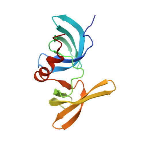 | |
UniProt & NIH Common Fund Data Resources | |||||
PHAROS: Q12888 GTEx: ENSG00000067369 | |||||
Entity Groups | |||||
| Sequence Clusters | 30% Identity50% Identity70% Identity90% Identity95% Identity100% Identity | ||||
| UniProt Group | Q12888 | ||||
Sequence AnnotationsExpand | |||||
Reference Sequence | |||||
| Ligands 3 Unique | |||||
|---|---|---|---|---|---|
| ID | Chains | Name / Formula / InChI Key | 2D Diagram | 3D Interactions | |
| WNQ (Subject of Investigation/LOI) Download:Ideal Coordinates CCD File | C [auth A], G [auth B] | 4-(4-methylpiperazine-1-sulfonyl)benzamide C12 H17 N3 O3 S DGYBGNBRDCUNEP-UHFFFAOYSA-N |  | ||
| SO4 Download:Ideal Coordinates CCD File | H [auth B], I [auth B] | SULFATE ION O4 S QAOWNCQODCNURD-UHFFFAOYSA-L |  | ||
| UNX Download:Ideal Coordinates CCD File | D [auth A], E [auth A], F [auth A] | UNKNOWN ATOM OR ION X |  | ||
| Length ( Å ) | Angle ( ˚ ) |
|---|---|
| a = 43.523 | α = 90 |
| b = 51.188 | β = 90 |
| c = 105.601 | γ = 90 |
| Software Name | Purpose |
|---|---|
| SCALEPACK | data scaling |
| PHASER | phasing |
| REFMAC | refinement |
| PDB_EXTRACT | data extraction |
| HKL-3000 | data reduction |
| Funding Organization | Location | Grant Number |
|---|---|---|
| Other private | -- |


