Download Mendeley
Proposal of metagenomic-origin LRA-5 as a precursor of active beta-lactamases through Tyr69Gln and Val166Glu amino acid substitutions: a functional and structural analysis.
D'Amico Gonzalez, G., Rodriguez, M.M., Penzotti, P., Brunetti, F., Ghiglione, B., Moe, L.A., Centron, D., Gutkind, G., Gao, L., Haider, S., Powers, R.A., Klinke, S., Power, P.(2026) Antimicrob Agents Chemother 70: e0067525-e0067525
- PubMed: 41312992 Search on PubMedSearch on PubMed Central
- DOI: https://doi.org/10.1128/aac.00675-25
- Primary Citation Related Structures:
8EO5 - PubMed Abstract:
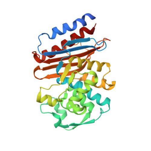
Wild-type LRA-5, recovered from Alaskan soil samples, shares no more than 33% amino acid sequence identity with enzymes from pathogens like PER β-lactamases. Recombinant E. coli expressing wild-type LRA-5 and its engineered variants LRA-5 Y69Q and LRA-5 V166E showed MIC values equivalent to control strains. However, LRA-5 Y69Q/V166E displayed MICs above the resistant breakpoint for some β-lactams. Kinetic parameters correlated with the MICs, showing that the catalytic efficiency of LRA-5 Y69Q/V166E was comparable to those from class A β-lactamases, such as CTX-M-15, PER-2, and KPC-2. LRA-5 Y69Q/V166E exhibited k cat / K m values up to 11,000-fold higher compared to wild-type LRA-5, which is associated with the presence of Glu166. The X-ray crystallographic structure of wild-type LRA-5 (1.80 Å; PDB 8EO5) shows that the lack of both Glu166 and a deacylation water molecule contributes to a biologically insignificant activity. Interactions observed between LRA-5 and ceftazidime (2.35 Å; PDB 8EO6) show structural conservation with other β-lactamases. In contrast, the crystallographic structure of LRA-5 Y69Q/V166E (2.15 Å; PDB 8EO7) bears a deacylation water molecule that is associated with the increase in catalytic activity compared to the wild-type variant. Circular dichroism results confirm that amino acid substitutions in LRA-5 do not affect the overall content of the secondary/tertiary structures. Evidence suggests that alternative evolutionary paths could have occurred for β-lactamases like LRA-5, produced by environmental microorganisms: (i) proteins having similar structural features than active β-lactamases may accumulate a small number of mutations (e.g., Y69Q/V166E) to yield active enzymes and (ii) the β-lactamase fold may have lost key residues in the absence of antibiotics.
- Facultad de Farmacia y Bioquímica, Universidad de Buenos Aires, Instituto de Investigaciones en Bacteriología y Virología Molecular (IBaViM), Buenos Aires, Argentina.
Organizational Affiliation: