8EMW

Phospholipase C beta 3 (PLCb3) in complex with Gbg on liposomes

  • Classification: HYDROLASE
  • Organism(s): Homo sapiens
  • Expression System: Trichoplusia ni
  • Mutation(s): No 
  • Membrane Protein: Yes  mpstruc

  • Deposited: 2022-09-28 Released: 2023-05-24 
  • Deposition Author(s): Falzone, M.E., MacKinnon, R.
  • Funding Organization(s): Howard Hughes Medical Institute (HHMI), National Institutes of Health/National Institute of General Medical Sciences (NIH/NIGMS)

Experimental Data Snapshot

  • Method: ELECTRON MICROSCOPY
  • Resolution: 3.50 Å
  • Aggregation State: PARTICLE 
  • Reconstruction Method: SINGLE PARTICLE 

wwPDB Validation   3D Report Full Report


This is version 1.0 of the entry. See complete history


Literature

G beta gamma activates PIP2 hydrolysis by recruiting and orienting PLC beta on the membrane surface.

Falzone, M.E.MacKinnon, R.

(2023) Proc Natl Acad Sci U S A 120: e2301121120-e2301121120

  • DOI: https://doi.org/10.1073/pnas.2301121120
  • Primary Citation of Related Structures:  
    8EMV, 8EMW, 8EMX

  • PubMed Abstract: 

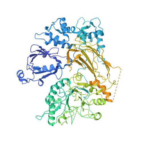
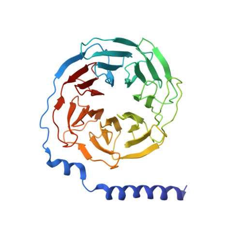
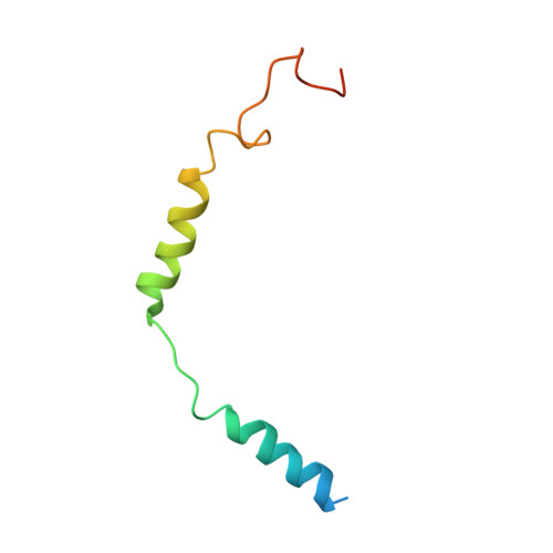
    Phospholipase C-βs (PLCβs) catalyze the hydrolysis of phosphatidylinositol 4, 5-bisphosphate [Formula: see text] into [Formula: see text] [Formula: see text] and [Formula: see text]  [Formula: see text]. [Formula: see text] regulates the activity of many membrane proteins, while IP3 and DAG lead to increased intracellular Ca 2+ levels and activate protein kinase C, respectively. PLCβs are regulated by G protein-coupled receptors through direct interaction with [Formula: see text] and [Formula: see text] and are aqueous-soluble enzymes that must bind to the cell membrane to act on their lipid substrate. This study addresses the mechanism by which [Formula: see text] activates PLCβ 3. We show that PLCβ 3 functions as a slow Michaelis-Menten enzyme ( [Formula: see text] ) on membrane surfaces. We used membrane partitioning experiments to study the solution-membrane localization equilibrium of PLCβ 3. Its partition coefficient is such that only a small quantity of  PLCβ 3 exists in the membrane in the absence of [Formula: see text] . When [Formula: see text] is present, equilibrium binding on the membrane surface increases PLCβ 3 in the membrane, increasing [Formula: see text] in proportion. Atomic structures on membrane vesicle surfaces show that two [Formula: see text] anchor  PLCβ 3 with its catalytic site oriented toward the membrane surface. Taken together, the enzyme kinetic, membrane partitioning, and structural data show that [Formula: see text] activates PLCβ by increasing its concentration on the membrane surface and orienting its catalytic core to engage [Formula: see text] . This principle of activation explains rapid stimulated catalysis with low background activity, which is essential to the biological processes mediated by [Formula: see text] , IP3 , and DAG .


  • Organizational Affiliation

    Laboratory of Molecular Neurobiology and Biophysics, The Rockefeller University, New York, NY 10065.


Macromolecules
Find similar proteins by:  (by identity cutoff)  |  3D Structure
Entity ID: 1
MoleculeChains Sequence LengthOrganismDetailsImage
1-phosphatidylinositol 4,5-bisphosphate phosphodiesterase beta-31,232Homo sapiensMutation(s): 0 
Gene Names: PLCB3
EC: 3.1.4.11
Membrane Entity: Yes 
UniProt & NIH Common Fund Data Resources
Find proteins for Q01970 (Homo sapiens)
Explore Q01970 
Go to UniProtKB:  Q01970
PHAROS:  Q01970
GTEx:  ENSG00000149782 
Entity Groups  
Sequence Clusters30% Identity50% Identity70% Identity90% Identity95% Identity100% Identity
UniProt GroupQ01970
Sequence Annotations
Expand
  • Reference Sequence
Find similar proteins by:  (by identity cutoff)  |  3D Structure
Entity ID: 2
MoleculeChains Sequence LengthOrganismDetailsImage
Guanine nucleotide-binding protein G(I)/G(S)/G(T) subunit beta-1B,
D [auth C]
340Homo sapiensMutation(s): 0 
Gene Names: GNB1
UniProt & NIH Common Fund Data Resources
Find proteins for P62873 (Homo sapiens)
Explore P62873 
Go to UniProtKB:  P62873
PHAROS:  P62873
GTEx:  ENSG00000078369 
Entity Groups  
Sequence Clusters30% Identity50% Identity70% Identity90% Identity95% Identity100% Identity
UniProt GroupP62873
Sequence Annotations
Expand
  • Reference Sequence
Find similar proteins by:  (by identity cutoff)  |  3D Structure
Entity ID: 3
MoleculeChains Sequence LengthOrganismDetailsImage
Guanine nucleotide-binding protein G(I)/G(S)/G(O) subunit gamma-2C [auth G],
E [auth D]
70Homo sapiensMutation(s): 0 
Gene Names: GNG2
Membrane Entity: Yes 
UniProt & NIH Common Fund Data Resources
Find proteins for P59768 (Homo sapiens)
Explore P59768 
Go to UniProtKB:  P59768
PHAROS:  P59768
GTEx:  ENSG00000186469 
Entity Groups  
Sequence Clusters30% Identity50% Identity70% Identity90% Identity95% Identity100% Identity
UniProt GroupP59768
Sequence Annotations
Expand
  • Reference Sequence
Small Molecules
Ligands 1 Unique
IDChains Name / Formula / InChI Key2D Diagram3D Interactions
CA
Query on CA

Download Ideal Coordinates CCD File 
F [auth A]CALCIUM ION
Ca
BHPQYMZQTOCNFJ-UHFFFAOYSA-N
Experimental Data & Validation

Experimental Data

  • Method: ELECTRON MICROSCOPY
  • Resolution: 3.50 Å
  • Aggregation State: PARTICLE 
  • Reconstruction Method: SINGLE PARTICLE 
EM Software:
TaskSoftware PackageVersion
RECONSTRUCTIONcryoSPARC
MODEL REFINEMENTPHENIX

Structure Validation

View Full Validation Report


Entry History & Funding Information

Deposition Data


Funding OrganizationLocationGrant Number
Howard Hughes Medical Institute (HHMI)United States--
National Institutes of Health/National Institute of General Medical Sciences (NIH/NIGMS)United StatesF32GM142137

Revision History  (Full details and data files)

  • Version 1.0: 2023-05-24
    Type: Initial release