8EM5 | pdb_00008em5

Mycobacterium thermoresistible MmpS5


Experimental Data Snapshot

  • Method: X-RAY DIFFRACTION
  • Resolution: 1.95 Å
  • R-Value Free: 
    0.194 (Depositor), 0.193 (DCC) 
  • R-Value Work: 
    0.161 (Depositor), 0.161 (DCC) 
  • R-Value Observed: 
    0.165 (Depositor) 

wwPDB Validation 3D Report Full Report

Validation slider image for 8EM5

This is version 1.2 of the entry. See complete history

Literature

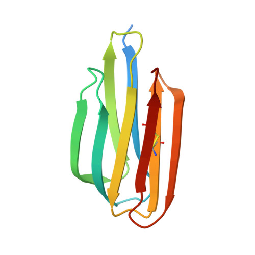
The structure of Mycobacterium thermoresistibile MmpS5 reveals a conserved disulfide bond across mycobacteria.

Cuthbert, B.J.Mendoza, J.de Miranda, R.Papavinasasundaram, K.Sassetti, C.M.Goulding, C.W.

(2024) Metallomics 16

  • DOI: https://doi.org/10.1093/mtomcs/mfae011
  • Primary Citation Related Structures: 
    8EM5

  • PubMed Abstract: 

    The tuberculosis (TB) emergency has been a pressing health threat for decades. With the emergence of drug-resistant TB and complications from the COVID-19 pandemic, the TB health crisis is more serious than ever. Mycobacterium tuberculosis (Mtb), the causative agent of TB, requires iron for its survival. Thus, Mtb has evolved several mechanisms to acquire iron from the host. Mtb produces two siderophores, mycobactin and carboxymycobactin, which scavenge for host iron. Mtb siderophore-dependent iron acquisition requires the export of apo-siderophores from the cytosol to the host environment and import of iron-bound siderophores. The export of Mtb apo-siderophores across the inner membrane is facilitated by two mycobacterial inner membrane proteins with their cognate periplasmic accessory proteins, designated MmpL4/MmpS4 and MmpL5/MmpS5. Notably, the Mtb MmpL4/MmpS4 and MmpL5/MmpS5 complexes have also been implicated in the efflux of anti-TB drugs. Herein, we solved the crystal structure of M. thermoresistibile MmpS5. The MmpS5 structure reveals a previously uncharacterized, biologically relevant disulfide bond that appears to be conserved across the Mycobacterium MmpS4/S5 homologs, and comparison with structural homologs suggests that MmpS5 may be dimeric.


  • Organizational Affiliation
    • Department of Molecular Biology & Biochemistry, University of California Irvine, Irvine, CA 92697, USA.

Macromolecule Content 

  • Total Structure Weight: 72.33 kDa 
  • Atom Count: 4,986 
  • Modeled Residue Count: 552 
  • Deposited Residue Count: 618 
  • Unique protein chains: 1

Macromolecules

Find similar proteins by:|  3D Structure
Entity ID: 1
MoleculeChains  Sequence LengthOrganismDetailsImage
MmpS5
A, B, C, D, E
A, B, C, D, E, F
103Mycolicibacterium thermoresistibileMutation(s): 0 
Gene Names: KEK_05752
UniProt
Find proteins for G7CDU2 (Mycolicibacterium thermoresistibile (strain ATCC 19527 / DSM 44167 / CIP 105390 / JCM 6362 / NCTC 10409 / 316))
Explore G7CDU2 
Go to UniProtKB:  G7CDU2
Entity Groups
Sequence Clusters30% Identity50% Identity70% Identity90% Identity95% Identity100% Identity
UniProt GroupG7CDU2
Sequence Annotations
Expand
Reference Sequence

Small Molecules

Ligands 4 Unique
IDChains Name / Formula / InChI Key2D Diagram3D Interactions
12P

Query on 12P


Download:Ideal Coordinates CCD File
G [auth A]
GA [auth E]
O [auth B]
Q [auth B]
S [auth C]
G [auth A],
GA [auth E],
O [auth B],
Q [auth B],
S [auth C],
T [auth C],
X [auth D]
DODECAETHYLENE GLYCOL
C24 H50 O13
WRZXKWFJEFFURH-UHFFFAOYSA-N
1PE

Query on 1PE


Download:Ideal Coordinates CCD File
K [auth A]PENTAETHYLENE GLYCOL
C10 H22 O6
JLFNLZLINWHATN-UHFFFAOYSA-N
IOD

Query on IOD


Download:Ideal Coordinates CCD File
AA [auth D]
IA [auth F]
JA [auth F]
KA [auth F]
LA [auth F]
AA [auth D],
IA [auth F],
JA [auth F],
KA [auth F],
LA [auth F],
U [auth C],
V [auth C],
Z [auth D]
IODIDE ION
I
XMBWDFGMSWQBCA-UHFFFAOYSA-M
GOL

Query on GOL


Download:Ideal Coordinates CCD File
BA [auth E]
CA [auth E]
DA [auth E]
EA [auth E]
FA [auth E]
BA [auth E],
CA [auth E],
DA [auth E],
EA [auth E],
FA [auth E],
H [auth A],
HA [auth F],
I [auth A],
J [auth A],
L [auth A],
M [auth A],
N [auth B],
P [auth B],
R [auth C],
W [auth D],
Y [auth D]
GLYCEROL
C3 H8 O3
PEDCQBHIVMGVHV-UHFFFAOYSA-N

Experimental Data & Validation

Experimental Data

  • Method: X-RAY DIFFRACTION
  • Resolution: 1.95 Å
  • R-Value Free:  0.194 (Depositor), 0.193 (DCC) 
  • R-Value Work:  0.161 (Depositor), 0.161 (DCC) 
  • R-Value Observed: 0.165 (Depositor) 
Space Group: P 1 21 1
Unit Cell:
Length ( Å )Angle ( ˚ )
a = 50.284α = 90
b = 85.257β = 95.19
c = 89.124γ = 90
Software Package:
Software NamePurpose
PHENIXrefinement
XDSdata reduction
Aimlessdata scaling
AutoSolphasing

Structure Validation

View Full Validation Report


Entry History 

& Funding Information

Deposition Data


Funding OrganizationLocationGrant Number
National Institutes of Health/National Institute Of Allergy and Infectious Diseases (NIH/NIAID)United States--

Revision History  (Full details and data files)

  • Version 1.0: 2023-09-27
    Type: Initial release
  • Version 1.1: 2024-05-15
    Changes: Database references
  • Version 1.2: 2024-10-16
    Changes: Structure summary