


Crystal Structure of UDP-N-acetylmuramate-L-alanine ligase (UDP-N-acetylmuramoyl-L-alanine synthetase, MurC) Pseudomonas aeruginosa
Abendroth, J., Horanyi, P.S., Hill, P., Lorimer, D.D., Edwards, T.E.To be published.
Experimental Data Snapshot
Starting Model: experimental
View more details
Entity ID: 1 | |||||
|---|---|---|---|---|---|
| Molecule | Chains | Sequence Length | Organism | Details | Image |
| UDP-N-acetylmuramate--L-alanine ligase | 316 | Pseudomonas aeruginosa PAO1 | Mutation(s): 0 Gene Names: murC, PA4411 EC: 6.3.2.8 | 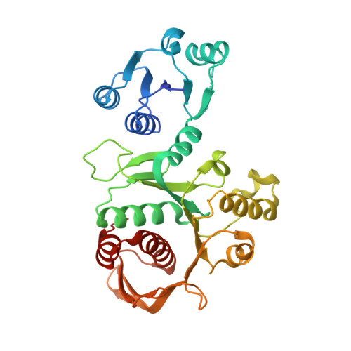 | |
UniProt | |||||
Entity Groups | |||||
| Sequence Clusters | 30% Identity50% Identity70% Identity90% Identity95% Identity100% Identity | ||||
| UniProt Group | Q9HW02 | ||||
Sequence AnnotationsExpand | |||||
Reference Sequence | |||||
| Ligands 2 Unique | |||||
|---|---|---|---|---|---|
| ID | Chains | Name / Formula / InChI Key | 2D Diagram | 3D Interactions | |
| WIU (Subject of Investigation/LOI) Download:Ideal Coordinates CCD File | I [auth A] K [auth B] M [auth C] O [auth D] Q [auth E] | (2R)-2-({4-[(5-tert-butyl-1-methyl-1H-pyrazol-3-yl)amino]-1H-pyrazolo[3,4-d]pyrimidin-6-yl}amino)-2-phenylethyl (2-aminoethyl)carbamate C24 H32 N10 O2 RUTNTFGYZLRUQX-KRWDZBQOSA-N |  | ||
| SO4 Download:Ideal Coordinates CCD File | J [auth A] L [auth B] N [auth C] P [auth D] R [auth E] | SULFATE ION O4 S QAOWNCQODCNURD-UHFFFAOYSA-L |  | ||
| Length ( Å ) | Angle ( ˚ ) |
|---|---|
| a = 285.26 | α = 90 |
| b = 109.2 | β = 112.403 |
| c = 108.71 | γ = 90 |
| Software Name | Purpose |
|---|---|
| XDS | data reduction |
| XSCALE | data scaling |
| PHENIX | refinement |
| PDB_EXTRACT | data extraction |
| PHASER | phasing |
| Funding Organization | Location | Grant Number |
|---|---|---|
| National Institutes of Health/National Institute Of Allergy and Infectious Diseases (NIH/NIAID) | United States | HHSN272201700059C |


