


Crystal structure of glutamate racemase from Helicobacter pylori in complex with a fragment
Cooling, G.T., Propp, J., Spies, M.A.To be published.
Experimental Data Snapshot
Starting Model: experimental
View more details
Entity ID: 1 | |||||
|---|---|---|---|---|---|
| Molecule | Chains | Sequence Length | Organism | Details | Image |
| Glutamate racemase | 260 | Helicobacter pylori | Mutation(s): 0 Gene Names: murI, OUM_0701 EC: 5.1.1.3 | 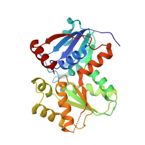 | |
UniProt | |||||
Entity Groups | |||||
| Sequence Clusters | 30% Identity50% Identity70% Identity90% Identity95% Identity100% Identity | ||||
| UniProt Group | K2K6A3 | ||||
Sequence AnnotationsExpand | |||||
Reference Sequence | |||||
| Ligands 5 Unique | |||||
|---|---|---|---|---|---|
| ID | Chains | Name / Formula / InChI Key | 2D Diagram | 3D Interactions | |
| WFI (Subject of Investigation/LOI) Download:Ideal Coordinates CCD File | F [auth A], I [auth B] | 1-[4-methyl-2-(pyridin-4-yl)-1,3-thiazol-5-yl]methanamine C10 H11 N3 S HSWRYUZXDMQDGC-UHFFFAOYSA-N |  | ||
| DGL Download:Ideal Coordinates CCD File | G [auth A], J [auth B] | D-GLUTAMIC ACID C5 H9 N O4 WHUUTDBJXJRKMK-GSVOUGTGSA-N |  | ||
| GOL Download:Ideal Coordinates CCD File | C [auth A], H [auth B] | GLYCEROL C3 H8 O3 PEDCQBHIVMGVHV-UHFFFAOYSA-N |  | ||
| DMS Download:Ideal Coordinates CCD File | D [auth A], E [auth A] | DIMETHYL SULFOXIDE C2 H6 O S IAZDPXIOMUYVGZ-UHFFFAOYSA-N |  | ||
| CL Download:Ideal Coordinates CCD File | K [auth B] | CHLORIDE ION Cl VEXZGXHMUGYJMC-UHFFFAOYSA-M |  | ||
| Length ( Å ) | Angle ( ˚ ) |
|---|---|
| a = 53.002 | α = 90 |
| b = 95.985 | β = 112.97 |
| c = 57.074 | γ = 90 |
| Software Name | Purpose |
|---|---|
| Aimless | data scaling |
| PHENIX | refinement |
| PDB_EXTRACT | data extraction |
| XDS | data reduction |
| PHASER | phasing |
| Funding Organization | Location | Grant Number |
|---|---|---|
| National Institutes of Health/National Institute of General Medical Sciences (NIH/NIGMS) | United States | RO1 GM097373 |


