8EB0

RNF216/E2-Ub/Ub transthiolation complex


Experimental Data Snapshot

  • Method: X-RAY DIFFRACTION
  • Resolution: 3.03 Å
  • R-Value Free: 0.339 
  • R-Value Work: 0.303 
  • R-Value Observed: 0.306 

wwPDB Validation   3D Report Full Report


This is version 1.1 of the entry. See complete history


Literature

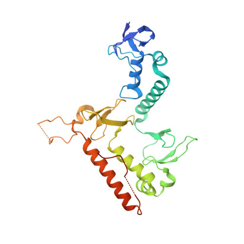
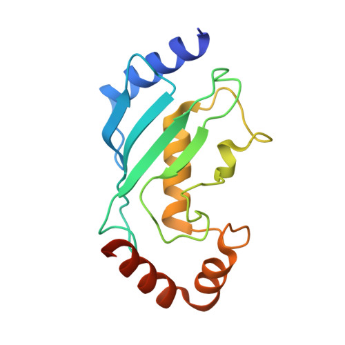
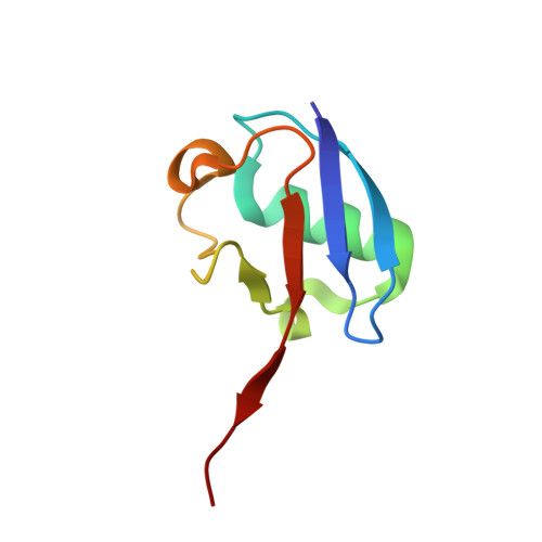
The unifying catalytic mechanism of the RING-between-RING E3 ubiquitin ligase family.

Wang, X.S.Cotton, T.R.Trevelyan, S.J.Richardson, L.W.Lee, W.T.Silke, J.Lechtenberg, B.C.

(2023) Nat Commun 14: 168-168

  • DOI: https://doi.org/10.1038/s41467-023-35871-z
  • Primary Citation of Related Structures:  
    8EAZ, 8EB0

  • PubMed Abstract: 

    The RING-between-RING (RBR) E3 ubiquitin ligase family in humans comprises 14 members and is defined by a two-step catalytic mechanism in which ubiquitin is first transferred from an E2 ubiquitin-conjugating enzyme to the RBR active site and then to the substrate. To define the core features of this catalytic mechanism, we here structurally and biochemically characterise the two RBRs HOIL-1 and RNF216. Crystal structures of both enzymes in their RBR/E2-Ub/Ub transthiolation complexes capturing the first catalytic step, together with complementary functional experiments, reveal the defining features of the RBR catalytic mechanism. RBRs catalyse ubiquitination via a conserved transthiolation complex structure that enables efficient E2-to-RBR ubiquitin transfer. Our data also highlight a conserved RBR allosteric activation mechanism by distinct ubiquitin linkages that suggests RBRs employ a feed-forward mechanism. We finally identify that the HOIL-1 RING2 domain contains an unusual Zn2/Cys6 binuclear cluster that is required for catalytic activity and substrate ubiquitination.


  • Organizational Affiliation

    Ubiquitin Signalling Division, The Walter and Eliza Hall Institute of Medical Research, Parkville, Victoria, 3052, Australia.


Macromolecules
Find similar proteins by:  (by identity cutoff)  |  3D Structure
Entity ID: 1
MoleculeChains Sequence LengthOrganismDetailsImage
E3 ubiquitin-protein ligase RNF216277Homo sapiensMutation(s): 1 
Gene Names: RNF216TRIAD3UBCE7IP1ZIN
UniProt & NIH Common Fund Data Resources
Find proteins for Q9NWF9 (Homo sapiens)
Explore Q9NWF9 
Go to UniProtKB:  Q9NWF9
PHAROS:  Q9NWF9
GTEx:  ENSG00000011275 
Entity Groups  
Sequence Clusters30% Identity50% Identity70% Identity90% Identity95% Identity100% Identity
UniProt GroupQ9NWF9
Sequence Annotations
Expand
  • Reference Sequence
Find similar proteins by:  (by identity cutoff)  |  3D Structure
Entity ID: 2
MoleculeChains Sequence LengthOrganismDetailsImage
Ubiquitin-conjugating enzyme E2 L3157Homo sapiensMutation(s): 1 
Gene Names: UBE2L3UBCE7UBCH7
UniProt & NIH Common Fund Data Resources
Find proteins for P68036 (Homo sapiens)
Explore P68036 
Go to UniProtKB:  P68036
PHAROS:  P68036
GTEx:  ENSG00000185651 
Entity Groups  
Sequence Clusters30% Identity50% Identity70% Identity90% Identity95% Identity100% Identity
UniProt GroupP68036
Sequence Annotations
Expand
  • Reference Sequence
Find similar proteins by:  (by identity cutoff)  |  3D Structure
Entity ID: 3
MoleculeChains Sequence LengthOrganismDetailsImage
Ubiquitin
C, D
76Homo sapiensMutation(s): 0 
Gene Names: UBC
UniProt & NIH Common Fund Data Resources
Find proteins for P0CG48 (Homo sapiens)
Explore P0CG48 
Go to UniProtKB:  P0CG48
PHAROS:  P0CG48
GTEx:  ENSG00000150991 
Entity Groups  
Sequence Clusters30% Identity50% Identity70% Identity90% Identity95% Identity100% Identity
UniProt GroupP0CG48
Sequence Annotations
Expand
  • Reference Sequence
Small Molecules
Ligands 2 Unique
IDChains Name / Formula / InChI Key2D Diagram3D Interactions
SO4
Query on SO4

Download Ideal Coordinates CCD File 
L [auth A],
M [auth D],
N [auth D]
SULFATE ION
O4 S
QAOWNCQODCNURD-UHFFFAOYSA-L
ZN
Query on ZN

Download Ideal Coordinates CCD File 
E [auth A]
F [auth A]
G [auth A]
H [auth A]
I [auth A]
E [auth A],
F [auth A],
G [auth A],
H [auth A],
I [auth A],
J [auth A],
K [auth A]
ZINC ION
Zn
PTFCDOFLOPIGGS-UHFFFAOYSA-N
Experimental Data & Validation

Experimental Data

  • Method: X-RAY DIFFRACTION
  • Resolution: 3.03 Å
  • R-Value Free: 0.339 
  • R-Value Work: 0.303 
  • R-Value Observed: 0.306 
  • Space Group: C 1 2 1
Unit Cell:
Length ( Å )Angle ( ˚ )
a = 152.545α = 90
b = 70.363β = 108.7
c = 65.152γ = 90
Software Package:
Software NamePurpose
PHENIXrefinement
XDSdata reduction
Aimlessdata scaling
PHASERphasing
Cootmodel building

Structure Validation

View Full Validation Report


Entry History & Funding Information

Deposition Data


Funding OrganizationLocationGrant Number
National Health and Medical Research Council (NHMRC, Australia)AustraliaGNT1182757

Revision History  (Full details and data files)

  • Version 1.0: 2023-01-18
    Type: Initial release
  • Version 1.1: 2023-10-25
    Changes: Data collection, Refinement description