8E7W | pdb_00008e7w

RsTSPO A139T with Heme


Experimental Data Snapshot

  • Method: X-RAY DIFFRACTION
  • Resolution: 2.10 Å
  • R-Value Free: 
    0.253 (Depositor), 0.252 (DCC) 
  • R-Value Work: 
    0.195 (Depositor), 0.195 (DCC) 
  • R-Value Observed: 
    0.197 (Depositor) 

Starting Model: experimental
View more details

wwPDB Validation 3D Report Full Report

Validation slider image for 8E7W

Ligand Structure Quality Assessment 


This is version 1.2 of the entry. See complete history

Literature

New TSPO Crystal Structures of Mutant and Heme-Bound Forms with Altered Flexibility, Ligand Binding, and Porphyrin Degradation Activity.

Liu, J.Hiser, C.Li, F.Hall, R.Garavito, R.M.Ferguson-Miller, S.

(2023) Biochemistry 62: 1262-1273

  • DOI: https://doi.org/10.1021/acs.biochem.2c00612
  • Primary Citation Related Structures: 
    8E7W, 8E7X, 8E7Y, 8E7Z

  • PubMed Abstract: 

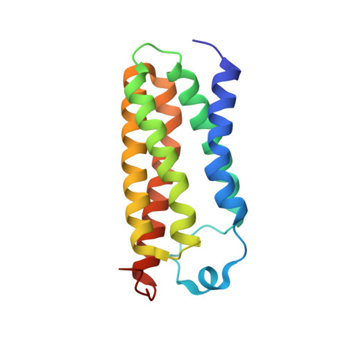
    The ancient protein TSPO (translocator protein 18kD) is found in all kingdoms and was originally identified as a binding site of benzodiazepine drugs. Its physiological function remains unclear, although porphyrins are conserved ligands. Several crystal structures of bacterial TSPO and nuclear magnetic resonance structures of a mouse form have revealed monomer and dimer configurations, but there have been no reports of structures with a physiological ligand. Here, we present the first X-ray structures of Rhodobacter sphaeroides TSPO with a physiological ligand bound. Two different variants (substituting threonine for alanine at position 139 (A139T) and phenylalanine for alanine at position 138 (A138F)) yielded well-diffracting crystals giving structures of both apo- and heme-containing forms. Both variants have wild-type micromolar affinity for heme and protoporphyrin IX, but A139T has very low ability to accelerate the breakdown of porphyrin in the presence of light and oxygen. The binding of heme to one protomer of the dimer of either mutant induces a more rigid structure, both in the heme-binding protomer and the protomer without heme bound, demonstrating an allosteric response. Ensemble refinement of the X-ray data reveals distinct regions of altered flexibility in response to single heme binding to the dimer. The A139T variant shows a more rigid structure overall, which may relate to extra hydrogen bonding of waters captured in the heme crevice. As TSPO has been suggested to have a role in heme delivery from mitochondria to the cytoplasm, the new structures provide potential clues regarding the structural basis of such activity.


  • Organizational Affiliation
    • Department of Chemistry, Michigan State University, East Lansing, Michigan 48824, United States.

Macromolecule Content 

  • Total Structure Weight: 61.11 kDa 
  • Atom Count: 4,178 
  • Modeled Residue Count: 462 
  • Deposited Residue Count: 474 
  • Unique protein chains: 1

Macromolecules

Find similar proteins by:|  3D Structure
Entity ID: 1
MoleculeChains  Sequence LengthOrganismDetailsImage
Tryptophan-rich sensory protein
A, B, C
158Cereibacter sphaeroidesMutation(s): 1 
Gene Names: tspOcrtK
Membrane Entity: Yes 
UniProt
Find proteins for Q9RFC8 (Cereibacter sphaeroides)
Explore Q9RFC8 
Go to UniProtKB:  Q9RFC8
Entity Groups
Sequence Clusters30% Identity50% Identity70% Identity90% Identity95% Identity100% Identity
UniProt GroupQ9RFC8
Sequence Annotations
Expand
Reference Sequence

Small Molecules

Ligands 3 Unique
IDChains Name / Formula / InChI Key2D Diagram3D Interactions
P6L

Query on P6L


Download:Ideal Coordinates CCD File
K [auth A](2S)-3-{[{[(2S)-2,3-DIHYDROXYPROPYL]OXY}(HYDROXY)PHOSPHORYL]OXY}-2-[(6E)-HEXADEC-6-ENOYLOXY]PROPYL (8E)-OCTADEC-8-ENOATE
C40 H75 O10 P
WLVNRDXLPAAELE-GDRYEXQRSA-N
HEM
(Subject of Investigation/LOI)

Query on HEM


Download:Ideal Coordinates CCD File
L [auth B]PROTOPORPHYRIN IX CONTAINING FE
C34 H32 Fe N4 O4
KABFMIBPWCXCRK-RGGAHWMASA-L
OLC

Query on OLC


Download:Ideal Coordinates CCD File
D [auth A]
E [auth A]
F [auth A]
G [auth A]
H [auth A]
D [auth A],
E [auth A],
F [auth A],
G [auth A],
H [auth A],
I [auth A],
J [auth A],
M [auth B],
N [auth B],
O [auth B],
P [auth B],
Q [auth B],
R [auth C],
S [auth C],
T [auth C],
U [auth C]
(2R)-2,3-dihydroxypropyl (9Z)-octadec-9-enoate
C21 H40 O4
RZRNAYUHWVFMIP-GDCKJWNLSA-N

Experimental Data & Validation

Experimental Data

  • Method: X-RAY DIFFRACTION
  • Resolution: 2.10 Å
  • R-Value Free:  0.253 (Depositor), 0.252 (DCC) 
  • R-Value Work:  0.195 (Depositor), 0.195 (DCC) 
  • R-Value Observed: 0.197 (Depositor) 
Space Group: C 1 2 1
Unit Cell:
Length ( Å )Angle ( ˚ )
a = 58.399α = 90
b = 99.568β = 100.16
c = 95.812γ = 90
Software Package:
Software NamePurpose
PHENIXrefinement
PDB_EXTRACTdata extraction
XDSdata reduction
SCALAdata scaling
PHASERphasing

Structure Validation

View Full Validation Report


Ligand Structure Quality Assessment 


Entry History 

& Funding Information

Deposition Data


Funding OrganizationLocationGrant Number
National Institutes of Health/National Institute of General Medical Sciences (NIH/NIGMS)United States--

Revision History  (Full details and data files)

  • Version 1.0: 2023-03-29
    Type: Initial release
  • Version 1.1: 2023-04-19
    Changes: Database references, Refinement description
  • Version 1.2: 2023-10-25
    Changes: Data collection, Refinement description