An African-Specific Variant of TP53 Reveals PADI4 as a Regulator of p53-Mediated Tumor Suppression.
Indeglia, A., Leung, J.C., Miller, S.A., Leu, J.I., Dougherty, J.F., Clarke, N.L., Kirven, N.A., Shao, C., Ke, L., Lovell, S., Barnoud, T., Lu, D.Y., Lin, C., Kannan, T., Battaile, K.P., Yang, T.H.L., Batista Oliva, I., Claiborne, D.T., Vogel, P., Liu, L., Liu, Q., Nefedova, Y., Cassel, J., Auslander, N., Kossenkov, A.V., Karanicolas, J., Murphy, M.E.(2023) Cancer Discov 13: 1696-1719
- PubMed: 37140445 Search on PubMedSearch on PubMed Central
- DOI: https://doi.org/10.1158/2159-8290.CD-22-1315
- Primary Citation Related Structures:
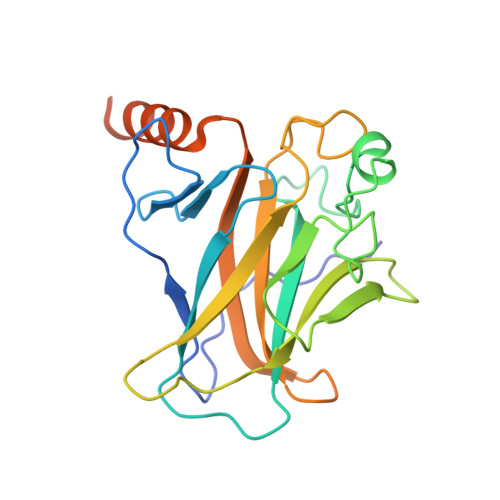
8E7A, 8E7B - PubMed Abstract:
TP53 is the most frequently mutated gene in cancer, yet key target genes for p53-mediated tumor suppression remain unidentified. Here, we characterize a rare, African-specific germline variant of TP53 in the DNA-binding domain Tyr107His (Y107H). Nuclear magnetic resonance and crystal structures reveal that Y107H is structurally similar to wild-type p53. Consistent with this, we find that Y107H can suppress tumor colony formation and is impaired for the transactivation of only a small subset of p53 target genes; this includes the epigenetic modifier PADI4, which deiminates arginine to the nonnatural amino acid citrulline. Surprisingly, we show that Y107H mice develop spontaneous cancers and metastases and that Y107H shows impaired tumor suppression in two other models. We show that PADI4 is itself tumor suppressive and that it requires an intact immune system for tumor suppression. We identify a p53-PADI4 gene signature that is predictive of survival and the efficacy of immune-checkpoint inhibitors. We analyze the African-centric Y107H hypomorphic variant and show that it confers increased cancer risk; we use Y107H in order to identify PADI4 as a key tumor-suppressive p53 target gene that contributes to an immune modulation signature and that is predictive of cancer survival and the success of immunotherapy. See related commentary by Bhatta and Cooks, p. 1518. This article is highlighted in the In This Issue feature, p. 1501.
- Program in Molecular and Cellular Oncogenesis, The Wistar Institute, Philadelphia, Pennsylvania.
Organizational Affiliation: