8E3I

CRYO-EM STRUCTURE OF the human MPSF IN COMPLEX WITH THE AUUAAA poly(A) signal


Experimental Data Snapshot

  • Method: ELECTRON MICROSCOPY
  • Resolution: 2.53 Å
  • Aggregation State: PARTICLE 
  • Reconstruction Method: SINGLE PARTICLE 

wwPDB Validation   3D Report Full Report


This is version 1.0 of the entry. See complete history


Literature

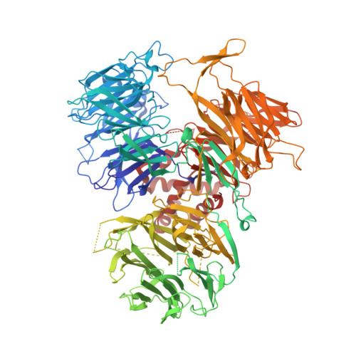
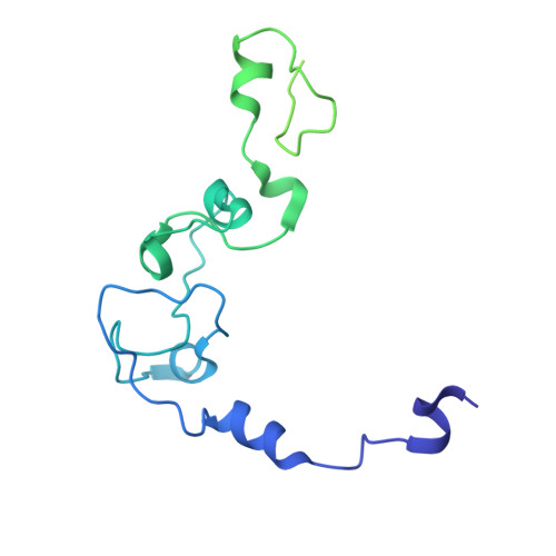
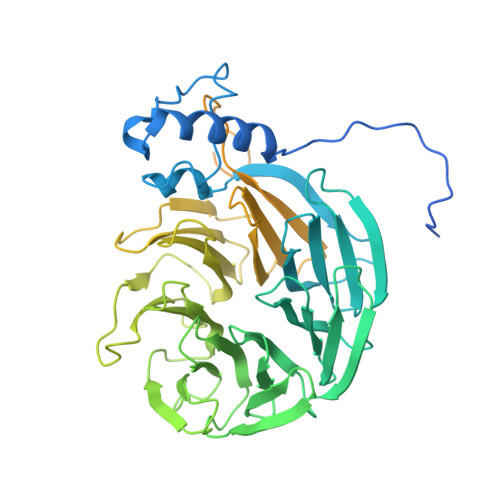
Molecular basis for the recognition of the AUUAAA polyadenylation signal by mPSF.

Gutierrez, P.A.Wei, J.Sun, Y.Tong, L.

(2022) RNA 28: 1534-1541

  • DOI: https://doi.org/10.1261/rna.079322.122
  • Primary Citation of Related Structures:  
    8E3I, 8E3Q

  • PubMed Abstract: 

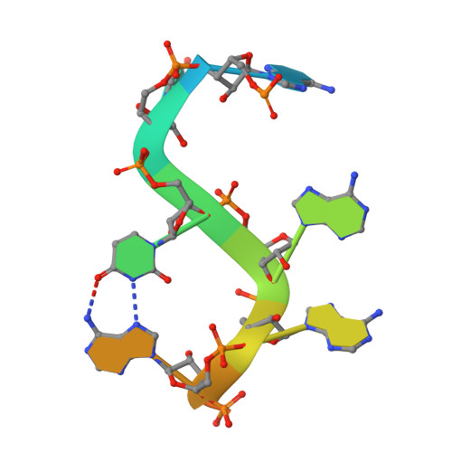
    The polyadenylation signal (PAS) is a key sequence element for 3'-end cleavage and polyadenylation of messenger RNA precursors (pre-mRNAs). This hexanucleotide motif is recognized by the mammalian polyadenylation specificity factor (mPSF), consisting of CPSF160, WDR33, CPSF30, and Fip1 subunits. Recent studies have revealed how the AAUAAA PAS, the most frequently observed PAS, is recognized by mPSF. We report here the structure of human mPSF in complex with the AUUAAA PAS, the second most frequently identified PAS. Conformational differences are observed for the A1 and U2 nucleotides in AUUAAA compared to the A1 and A2 nucleotides in AAUAAA, while the binding modes of the remaining 4 nt are essentially identical. The 5' phosphate of U2 moves by 2.6 Å and the U2 base is placed near the six-membered ring of A2 in AAUAAA, where it makes two hydrogen bonds with zinc finger 2 (ZF2) of CPSF30, which undergoes conformational changes as well. We also attempted to determine the binding modes of two rare PAS hexamers, AAGAAA and GAUAAA, but did not observe the RNA in the cryo-electron microscopy density. The residues in CPSF30 (ZF2 and ZF3) and WDR33 that recognize PAS are disordered in these two structures.


  • Organizational Affiliation

    Department of Biological Sciences, Columbia University, New York, New York 10027, USA.


Macromolecules

Find similar proteins by:  (by identity cutoff)  |  3D Structure
Entity ID: 1
MoleculeChains Sequence LengthOrganismDetailsImage
Cleavage and polyadenylation specificity factor subunit 11,443Homo sapiensMutation(s): 0 
Gene Names: CPSF1CPSF160
UniProt & NIH Common Fund Data Resources
Find proteins for Q10570 (Homo sapiens)
Explore Q10570 
Go to UniProtKB:  Q10570
PHAROS:  Q10570
GTEx:  ENSG00000071894 
Entity Groups  
Sequence Clusters30% Identity50% Identity70% Identity90% Identity95% Identity100% Identity
UniProt GroupQ10570
Sequence Annotations
Expand
  • Reference Sequence
Find similar proteins by:  (by identity cutoff)  |  3D Structure
Entity ID: 2
MoleculeChains Sequence LengthOrganismDetailsImage
Cleavage and polyadenylation specificity factor subunit 4B [auth C]245Homo sapiensMutation(s): 0 
Gene Names: CPSF4CPSF30NARNEB1
UniProt & NIH Common Fund Data Resources
Find proteins for O95639 (Homo sapiens)
Explore O95639 
Go to UniProtKB:  O95639
PHAROS:  O95639
GTEx:  ENSG00000160917 
Entity Groups  
Sequence Clusters30% Identity50% Identity70% Identity90% Identity95% Identity100% Identity
UniProt GroupO95639
Sequence Annotations
Expand
  • Reference Sequence
Find similar proteins by:  (by identity cutoff)  |  3D Structure
Entity ID: 4
MoleculeChains Sequence LengthOrganismDetailsImage
pre-mRNA 3' end processing protein WDR33D [auth B]572Homo sapiensMutation(s): 0 
Gene Names: WDR33WDC146
UniProt & NIH Common Fund Data Resources
Find proteins for Q9C0J8 (Homo sapiens)
Explore Q9C0J8 
Go to UniProtKB:  Q9C0J8
PHAROS:  Q9C0J8
GTEx:  ENSG00000136709 
Entity Groups  
Sequence Clusters30% Identity50% Identity70% Identity90% Identity95% Identity100% Identity
UniProt GroupQ9C0J8
Sequence Annotations
Expand
  • Reference Sequence

Find similar nucleic acids by:  Sequence   |   3D Structure  

Entity ID: 3
MoleculeChains LengthOrganismImage
RNA (5'-R(P*CP*AP*UP*UP*AP*AP*AP*CP*AP*AP*C)-3')C [auth E]11Homo sapiens
Sequence Annotations
Expand
  • Reference Sequence
Experimental Data & Validation

Experimental Data

  • Method: ELECTRON MICROSCOPY
  • Resolution: 2.53 Å
  • Aggregation State: PARTICLE 
  • Reconstruction Method: SINGLE PARTICLE 
EM Software:
TaskSoftware PackageVersion
MODEL REFINEMENTPHENIX

Structure Validation

View Full Validation Report


Entry History & Funding Information

Deposition Data


Funding OrganizationLocationGrant Number
National Institutes of Health/National Institute of General Medical Sciences (NIH/NIGMS)United StatesR35GM118093

Revision History  (Full details and data files)

  • Version 1.0: 2023-01-18
    Type: Initial release