8E3D

ZBTB7A Zinc Finger Domain Bound to DNA Duplex Containing CAST sequence (#11)


Experimental Data Snapshot

  • Method: X-RAY DIFFRACTION
  • Resolution: 2.62 Å
  • R-Value Free: 0.237 
  • R-Value Work: 0.206 
  • R-Value Observed: 0.207 

wwPDB Validation   3D Report Full Report


This is version 1.2 of the entry. See complete history


Literature

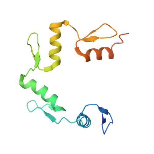
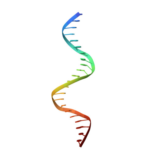
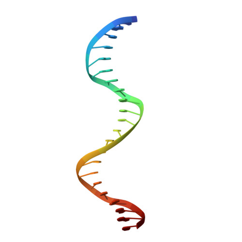
Structural basis for transcription factor ZBTB7A recognition of DNA and effects of ZBTB7A somatic mutations that occur in human acute myeloid leukemia.

Ren, R.Horton, J.R.Chen, Q.Yang, J.Liu, B.Huang, Y.Blumenthal, R.M.Zhang, X.Cheng, X.

(2023) J Biol Chem 299: 102885-102885

  • DOI: https://doi.org/10.1016/j.jbc.2023.102885
  • Primary Citation of Related Structures:  
    7N5U, 7N5V, 7N5W, 8E3D, 8E3E

  • PubMed Abstract: 

    ZBTB7A belongs to a small family of transcription factors having three members in humans (7A, 7B, and 7C). They share a BTB/POZ protein interaction domain at the amino end and a zinc-finger DNA-binding domain at the carboxyl end. They control the transcription of a wide range of genes, having varied functions in hematopoiesis, oncogenesis, and metabolism (in particular glycolysis). ZBTB7A-binding profiles at gene promoters contain a consensus G(a/c)CCC motif, followed by a CCCC sequence in some instances. Structural and mutational investigations suggest that DNA-specific contacts with the four-finger tandem array of ZBTB7A are formed sequentially, initiated from ZF1-ZF2 binding to G(a/c)CCC before spreading to ZF3-ZF4, which bind the DNA backbone and the 3' CCCC sequence, respectively. Here, we studied some mutations found in t(8;21)-positive acute myeloid leukemia patients that occur within the ZBTB7A DNA-binding domain. We determined that these mutations generally impair ZBTB7A DNA binding, with the most severe disruptions resulting from mutations in ZF1 and ZF2, and the least from a frameshift mutation in ZF3 that results in partial mislocalization. Information provided here on ZBTB7A-DNA interactions is likely applicable to ZBTB7B/C, which have overlapping functions with ZBTB7A in controlling primary metabolism.


  • Organizational Affiliation

    Department of Epigenetics and Molecular Carcinogenesis, University of Texas MD Anderson Cancer Center, Houston, Texas, USA.


Macromolecules

Find similar proteins by:  (by identity cutoff)  |  3D Structure
Entity ID: 1
MoleculeChains Sequence LengthOrganismDetailsImage
Zinc finger and BTB domain-containing protein 7AA,
D [auth B],
G [auth F]
133Homo sapiensMutation(s): 0 
Gene Names: ZBTB7AFBI1LRFZBTB7ZNF857A
UniProt & NIH Common Fund Data Resources
Find proteins for O95365 (Homo sapiens)
Explore O95365 
Go to UniProtKB:  O95365
PHAROS:  O95365
GTEx:  ENSG00000178951 
Entity Groups  
Sequence Clusters30% Identity50% Identity70% Identity90% Identity95% Identity100% Identity
UniProt GroupO95365
Sequence Annotations
Expand
  • Reference Sequence

Find similar nucleic acids by:  Sequence   |   3D Structure  

Entity ID: 2
MoleculeChains LengthOrganismImage
DNA (5'-D(*AP*TP*TP*TP*GP*GP*GP*GP*AP*GP*GP*GP*GP*TP*CP*TP*TP*TP*AP*AP*CP*C)-3')B [auth X],
E [auth D],
H
22Homo sapiens
Sequence Annotations
Expand
  • Reference Sequence

Find similar nucleic acids by:  Sequence   |   3D Structure  

Entity ID: 3
MoleculeChains LengthOrganismImage
DNA (5'-D(P*GP*GP*TP*AP*AP*AP*AP*GP*AP*CP*CP*CP*CP*TP*CP*CP*CP*CP*AP*AP*AP*T)-3')C [auth Y],
F [auth E],
I
22Homo sapiens
Sequence Annotations
Expand
  • Reference Sequence
Small Molecules
Ligands 2 Unique
IDChains Name / Formula / InChI Key2D Diagram3D Interactions
ZN
Query on ZN

Download Ideal Coordinates CCD File 
L [auth A]
M [auth A]
N [auth A]
O [auth A]
R [auth B]
L [auth A],
M [auth A],
N [auth A],
O [auth A],
R [auth B],
S [auth B],
T [auth B],
U [auth B],
W [auth F],
X [auth F],
Y [auth F],
Z [auth F]
ZINC ION
Zn
PTFCDOFLOPIGGS-UHFFFAOYSA-N
EDO
Query on EDO

Download Ideal Coordinates CCD File 
J [auth A],
K [auth A],
P [auth Y],
Q [auth B],
V [auth D]
1,2-ETHANEDIOL
C2 H6 O2
LYCAIKOWRPUZTN-UHFFFAOYSA-N
Experimental Data & Validation

Experimental Data

  • Method: X-RAY DIFFRACTION
  • Resolution: 2.62 Å
  • R-Value Free: 0.237 
  • R-Value Work: 0.206 
  • R-Value Observed: 0.207 
  • Space Group: P 6
Unit Cell:
Length ( Å )Angle ( ˚ )
a = 196.181α = 90
b = 196.181β = 90
c = 54.964γ = 120
Software Package:
Software NamePurpose
PHENIXrefinement
SERGUIdata collection
HKL-2000data reduction
HKL-2000data scaling
PHENIXphasing

Structure Validation

View Full Validation Report


Entry History & Funding Information

Deposition Data


Funding OrganizationLocationGrant Number
National Institutes of Health/National Institute of General Medical Sciences (NIH/NIGMS)United StatesR35GM134744
Cancer Prevention and Research Institute of Texas (CPRIT)United StatesRR160029

Revision History  (Full details and data files)

  • Version 1.0: 2023-02-08
    Type: Initial release
  • Version 1.1: 2023-02-15
    Changes: Database references
  • Version 1.2: 2023-10-25
    Changes: Data collection, Refinement description