8E1W | pdb_00008e1w

Neutron crystal structure of Panus similis AA9A at room temperature


Experimental Data Snapshot

  • Method: X-RAY DIFFRACTION
  • Resolution: 2.10 Å
  • R-Value Free: 
    0.170 (Depositor), 0.170 (DCC) 
  • R-Value Work: 
    0.148 (Depositor), 0.150 (DCC) 
  • R-Value Observed: 
    0.150 (Depositor) 

  • Method: NEUTRON DIFFRACTION
  • Resolution: 2.80 Å
  • R-Value Free: 
    0.260 (Depositor) 
  • R-Value Work: 
    0.225 (Depositor) 

Starting Model: experimental
View more details

wwPDB Validation   3D Report Full Report


This is version 2.1 of the entry. See complete history


Literature

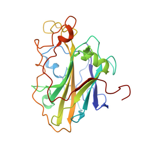
Joint X-ray/neutron structure of Lentinus similis AA9_A at room temperature.

Tandrup, T.Lo Leggio, L.Meilleur, F.

(2023) Acta Crystallogr F Struct Biol Commun 79: 1-7

  • DOI: https://doi.org/10.1107/S2053230X22011335
  • Primary Citation of Related Structures:  
    8E1W

  • PubMed Abstract: 

    Lytic polysaccharide monooxygenases (LPMOs) are copper metalloenzymes which cleave polysaccharides oxidatively and are important in pathogen biology, carbon cycling and biotechnology. The Lentinus similis family AA9 isoform A (LsAA9_A) has been extensively studied as a model system because its activity towards smaller soluble saccharide substrates has allowed detailed structural characterization of its interaction with a variety of substrates by X-ray crystallography at high resolution. Here, the joint X-ray/neutron room-temperature crystallographic structure of carbohydrate-free LsAA9_A in the copper(II) resting state refined against X-ray and neutron data at 2.1 and 2.8 Å resolution, respectively, is presented. The results provide an experimental determination of the protonation states of the copper(II)-coordinating residues and second-shell residues in LsAA9_A, paving the way for future neutron crystallographic studies of LPMO-carbohydrate complexes.


  • Organizational Affiliation
    • Department of Chemistry, University of Copenhagen, Universitetsparken 5, 2100 Copenhagen, Denmark.

Macromolecules
Find similar proteins by:  (by identity cutoff)  |  3D Structure
Entity ID: 1
MoleculeChains Sequence LengthOrganismDetailsImage
Endo-beta-1,4-glucanase D235Panus similisMutation(s): 0 
EC: 3.2.1.4 (PDB Primary Data), 1.14.99.56 (UniProt)
UniProt
Find proteins for A0A0S2GKZ1 (Panus similis)
Explore A0A0S2GKZ1 
Go to UniProtKB:  A0A0S2GKZ1
Entity Groups  
Sequence Clusters30% Identity50% Identity70% Identity90% Identity95% Identity100% Identity
UniProt GroupA0A0S2GKZ1
Glycosylation
Glycosylation Sites: 1
Sequence Annotations
Expand
  • Reference Sequence
Small Molecules
Modified Residues  1 Unique
IDChains TypeFormula2D DiagramParent
HIC
Query on HIC
A
L-PEPTIDE LINKINGC7 H11 N3 O2HIS
Experimental Data & Validation

Experimental Data

  • Method: X-RAY DIFFRACTION
  • Resolution: 2.10 Å
  • R-Value Free:  0.170 (Depositor), 0.170 (DCC) 
  • R-Value Work:  0.148 (Depositor), 0.150 (DCC) 
  • R-Value Observed: 0.150 (Depositor) 
  • Method: NEUTRON DIFFRACTION
  • Resolution: 2.80 Å
  • R-Value Free:  0.260 (Depositor) 
  • R-Value Work:  0.225 (Depositor) 
Space Group: P 41 3 2
Unit Cell:
Length ( Å )Angle ( ˚ )
a = 126.469α = 90
b = 126.469β = 90
c = 126.469γ = 90
Software Package:
Software NamePurpose
PHENIXrefinement
Mantiddata reduction
LAUENORMdata scaling
PHASERphasing

Structure Validation

View Full Validation Report


Entry History & Funding Information

Deposition Data


Funding OrganizationLocationGrant Number
Novo Nordisk FoundationDenmarkNNF17SA0027704
Danish Council for Independent ResearchDenmark8021-00273B
National Institute of Food and Agriculture (NIFA, United States)United StatesHatch211001

Revision History  (Full details and data files)

  • Version 1.0: 2023-01-11
    Type: Initial release
  • Version 2.0: 2023-01-18
    Changes: Atomic model
  • Version 2.1: 2024-04-03
    Changes: Data collection, Refinement description