8E1D | pdb_00008e1d

NMR-derived ensemble of the TAZ2 domain of p300 bound to the microphthalmia-associated transcription factor


Experimental Data Snapshot

  • Method: SOLUTION NMR
  • Conformers Calculated: 100 
  • Conformers Submitted: 20 
  • Selection Criteria: structures with the lowest energy 

wwPDB Validation 3D Report Full Report

Validation slider image for 8E1D

This is version 1.2 of the entry. See complete history

Literature

Structural basis of CBP/p300 recruitment by the microphthalmia-associated transcription factor.

Brown, A.D.Vergunst, K.L.Branch, M.Blair, C.M.Dupre, D.J.Baillie, G.S.Langelaan, D.N.

(2023) Biochim Biophys Acta Mol Cell Res 1870: 119520-119520

  • DOI: https://doi.org/10.1016/j.bbamcr.2023.119520
  • Primary Citation Related Structures: 
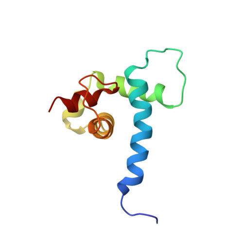
    8E1D

  • PubMed Abstract: 

    The microphthalmia-associated transcription factor (MITF) is a master regulator of the melanocyte cell lineage. Aberrant MITF activity can lead to multiple malignancies including skin cancer, where it modulates the progression and invasiveness of melanoma. MITF-regulated gene expression requires recruitment of the transcriptional co-regulator CBP/p300, but details of this process are not fully defined. In this study, we investigate the structural and functional interaction between the MITF N-terminal transactivation domain (MITF TAD ) and CBP/p300. Using pulldown assays and nuclear magnetic resonance spectroscopy we determined that MITF TAD is intrinsically disordered and binds to the TAZ1 and TAZ2 domains of CBP/p300 with moderate affinity. The solution-state structure of the MITF TAD :TAZ2 complex reveals that MITF interacts with a hydrophobic surface of TAZ2, while remaining somewhat dynamic. Peptide array and mutagenesis experiments determined that an acidic motif is integral to the MITF TAD :TAZ2 interaction and is necessary for transcriptional activity of MITF. Peptides that bind to the same surface of TAZ2 as MITF TAD , such as the adenoviral protein E1A, are capable of displacing MITF from TAZ2 and inhibiting transactivation. These findings provide insight into co-activator recruitment by MITF that are fundamental to our understanding of MITF targeted gene regulation and melanoma biology.


  • Organizational Affiliation
    • Department of Biochemistry & Molecular Biology, Dalhousie University, Halifax, NS B3H 4R2, Canada.

Macromolecule Content 

  • Total Structure Weight: 14.02 kDa 
  • Atom Count: 958 
  • Modeled Residue Count: 126 
  • Deposited Residue Count: 126 
  • Unique protein chains: 2

Macromolecules

Find similar proteins by:|  3D Structure
Entity ID: 1
MoleculeChains  Sequence LengthOrganismDetailsImage
Microphthalmia-associated transcription factorA [auth B]34Homo sapiensMutation(s): 0 
Gene Names: MITFBHLHE32
UniProt & NIH Common Fund Data Resources
Find proteins for O75030 (Homo sapiens)
Explore O75030 
Go to UniProtKB:  O75030
PHAROS:  O75030
GTEx:  ENSG00000187098 
Entity Groups
Sequence Clusters30% Identity50% Identity70% Identity90% Identity95% Identity100% Identity
UniProt GroupO75030
Sequence Annotations
Expand
Reference Sequence
Find similar proteins by:|  3D Structure
Entity ID: 2
MoleculeChains  Sequence LengthOrganismDetailsImage
Histone acetyltransferase p300B [auth A]92Homo sapiensMutation(s): 4 
Gene Names: EP300P300
EC: 2.3.1.48 (PDB Primary Data), 2.3.1 (PDB Primary Data)
UniProt & NIH Common Fund Data Resources
Find proteins for Q09472 (Homo sapiens)
Explore Q09472 
Go to UniProtKB:  Q09472
PHAROS:  Q09472
GTEx:  ENSG00000100393 
Entity Groups
Sequence Clusters30% Identity50% Identity70% Identity90% Identity95% Identity100% Identity
UniProt GroupQ09472
Sequence Annotations
Expand
Reference Sequence

Experimental Data & Validation

Experimental Data

  • Method: SOLUTION NMR
  • Conformers Calculated: 100 
  • Conformers Submitted: 20 
  • Selection Criteria: structures with the lowest energy 

Structure Validation

View Full Validation Report


Entry History 

& Funding Information

Deposition Data


Funding OrganizationLocationGrant Number
Other privateCanada--

Revision History  (Full details and data files)

  • Version 1.0: 2023-06-21
    Type: Initial release
  • Version 1.1: 2023-07-12
    Changes: Database references
  • Version 1.2: 2024-05-15
    Changes: Data collection, Database references