


Protic and Aprotic Interactions Systematically Perturbed and Mapped via MD and IR Spectroscopy
Kirsh, J.M., Kozuch, J., Weaver, J.B., Boxer, S.G.To be published.
Experimental Data Snapshot
Starting Model: experimental
View more details
Entity ID: 1 | |||||
|---|---|---|---|---|---|
| Molecule | Chains | Sequence Length | Organism | Details | Image |
| Photoactive yellow protein | 125 | Halorhodospira halophila | Mutation(s): 1 Gene Names: pyp | 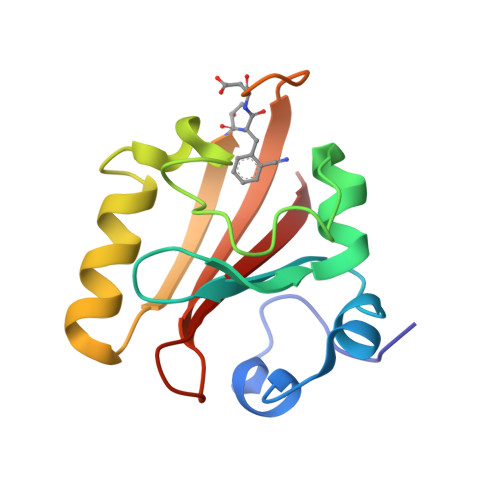 | |
UniProt | |||||
Entity Groups | |||||
| Sequence Clusters | 30% Identity50% Identity70% Identity90% Identity95% Identity100% Identity | ||||
| UniProt Group | P16113 | ||||
Sequence AnnotationsExpand | |||||
Reference Sequence | |||||
| Ligands 1 Unique | |||||
|---|---|---|---|---|---|
| ID | Chains | Name / Formula / InChI Key | 2D Diagram | 3D Interactions | |
| HC4 Download:Ideal Coordinates CCD File | B [auth A] | 4'-HYDROXYCINNAMIC ACID C9 H8 O3 NGSWKAQJJWESNS-ZZXKWVIFSA-N |  | ||
| Length ( Å ) | Angle ( ˚ ) |
|---|---|
| a = 66.305 | α = 90 |
| b = 66.305 | β = 90 |
| c = 40.692 | γ = 120 |
| Software Name | Purpose |
|---|---|
| PHENIX | refinement |
| XSCALE | data scaling |
| PDB_EXTRACT | data extraction |
| XDS | data reduction |
| PHENIX | phasing |
| Funding Organization | Location | Grant Number |
|---|---|---|
| National Institutes of Health/National Institute of General Medical Sciences (NIH/NIGMS) | United States | 2R35GM11804406 |
| National Science Foundation (NSF, United States) | United States | 1915727 |


