8DX0

VanSC CA domain

  • Classification: SIGNALING PROTEIN
  • Organism(s): Enterococcus
  • Expression System: Escherichia coli BL21(DE3)
  • Mutation(s): No 

  • Deposited: 2022-08-02 Released: 2023-03-22 
  • Deposition Author(s): Loll, P.J.
  • Funding Organization(s): National Institutes of Health/National Institute Of Allergy and Infectious Diseases (NIH/NIAID)

Experimental Data Snapshot

  • Method: X-RAY DIFFRACTION
  • Resolution: 1.45 Å
  • R-Value Free: 0.200 
  • R-Value Work: 0.185 
  • R-Value Observed: 0.186 

wwPDB Validation   3D Report Full Report


This is version 1.1 of the entry. See complete history


Literature

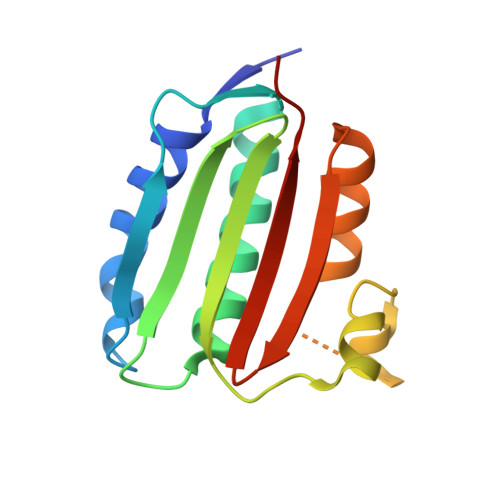
Structure of VanS from vancomycin-resistant enterococci: A sensor kinase with weak ATP binding.

Grasty, K.C.Guzik, C.D'Lauro, E.J.Padrick, S.B.Beld, J.Loll, P.J.

(2023) J Biol Chem 299: 103001-103001

  • DOI: https://doi.org/10.1016/j.jbc.2023.103001
  • Primary Citation of Related Structures:  
    8DVQ, 8DWZ, 8DX0

  • PubMed Abstract: 

    The VanRS two-component system regulates the resistance phenotype of vancomycin-resistant enterococci. VanS is a sensor histidine kinase that responds to the presence of vancomycin by autophosphorylating and subsequently transferring the phosphoryl group to the response regulator, VanR. The phosphotransfer activates VanR as a transcription factor, which initiates the expression of resistance genes. Structural information about VanS proteins has remained elusive, hindering the molecular-level understanding of their function. Here, we present X-ray crystal structures for the catalytic and ATP-binding (CA) domains of two VanS proteins, derived from vancomycin-resistant enterococci types A and C. Both proteins adopt the canonical Bergerat fold that has been observed for CA domains of other prokaryotic histidine kinases. We attempted to determine structures for the nucleotide-bound forms of both proteins; however, despite repeated efforts, these forms could not be crystallized, prompting us to measure the proteins' binding affinities for ATP. Unexpectedly, both CA domains displayed low affinities for the nucleotide, with K D values in the low millimolar range. Since these K D values are comparable to intracellular ATP concentrations, this weak substrate binding could reflect a way of regulating expression of the resistance phenotype.


  • Organizational Affiliation

    Department of Biochemistry and Molecular Biology, Drexel University College of Medicine, Philadelphia, Pennsylvania, USA.


Macromolecules
Find similar proteins by:  (by identity cutoff)  |  3D Structure
Entity ID: 1
MoleculeChains Sequence LengthOrganismDetailsImage
Histidine kinase
A, B
154EnterococcusMutation(s): 0 
Gene Names: vanSc
EC: 2.7.13.3
UniProt
Find proteins for Q9KJT3 (Enterococcus gallinarum)
Explore Q9KJT3 
Go to UniProtKB:  Q9KJT3
Entity Groups  
Sequence Clusters30% Identity50% Identity70% Identity90% Identity95% Identity100% Identity
UniProt GroupQ9KJT3
Sequence Annotations
Expand
  • Reference Sequence
Experimental Data & Validation

Experimental Data

  • Method: X-RAY DIFFRACTION
  • Resolution: 1.45 Å
  • R-Value Free: 0.200 
  • R-Value Work: 0.185 
  • R-Value Observed: 0.186 
  • Space Group: P 1 21 1
Unit Cell:
Length ( Å )Angle ( ˚ )
a = 62.7α = 90
b = 40.23β = 91.64
c = 64.71γ = 90
Software Package:
Software NamePurpose
PHENIXrefinement
XDSdata reduction
XSCALEdata scaling
PHASERphasing

Structure Validation

View Full Validation Report


Entry History & Funding Information

Deposition Data

  • Released Date: 2023-03-22 
  • Deposition Author(s): Loll, P.J.

Funding OrganizationLocationGrant Number
National Institutes of Health/National Institute Of Allergy and Infectious Diseases (NIH/NIAID)United StatesAI148679

Revision History  (Full details and data files)

  • Version 1.0: 2023-03-22
    Type: Initial release
  • Version 1.1: 2024-04-03
    Changes: Data collection, Refinement description