8DVL

Crystal structure of LRP6 E3E4 in complex with disulfide constrained peptide E3.18


Experimental Data Snapshot

  • Method: X-RAY DIFFRACTION
  • Resolution: 2.50 Å
  • R-Value Free: 0.245 
  • R-Value Work: 0.197 
  • R-Value Observed: 0.201 

wwPDB Validation   3D Report Full Report


Ligand Structure Quality Assessment 


This is version 1.3 of the entry. See complete history


Literature

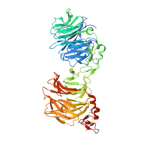
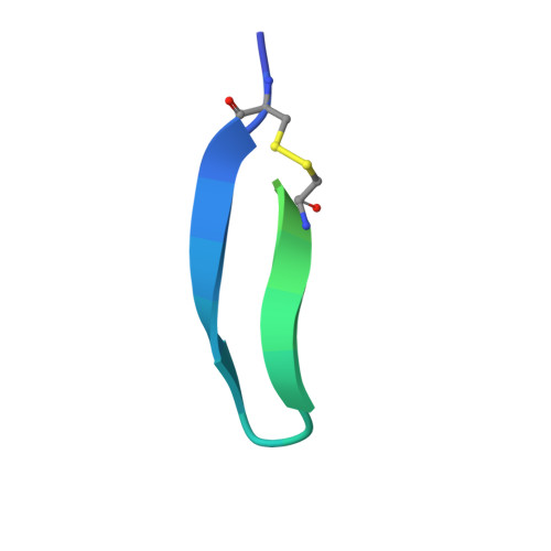
Synthetic Multivalent Disulfide-Constrained Peptide Agonists Potentiate Wnt1/ beta-Catenin Signaling via LRP6 Coreceptor Clustering.

Thakur, A.K.Miller, S.E.Liau, N.P.D.Hwang, S.Hansen, S.de Sousa E Melo, F.Sudhamsu, J.Hannoush, R.N.

(2023) ACS Chem Biol 18: 772-784

  • DOI: https://doi.org/10.1021/acschembio.2c00753
  • Primary Citation of Related Structures:  
    8DVL, 8DVM, 8DVN

  • PubMed Abstract: 

    Wnt ligands are critical for tissue homeostasis and form a complex with LRP6 and frizzled coreceptors to initiate Wnt/β-catenin signaling. Yet, how different Wnts achieve various levels of signaling activation through distinct domains on LRP6 remains elusive. Developing tool ligands that target individual LRP6 domains could help elucidate the mechanism of Wnt signaling regulation and uncover pharmacological approaches for pathway modulation. We employed directed evolution of a disulfide constrained peptide (DCP) to identify molecules that bind to the third β-propeller domain of LRP6. The DCPs antagonize Wnt3a while sparing Wnt1 signaling. Using PEG linkers with different geometries, we converted the Wnt3a antagonist DCPs to multivalent molecules that potentiated Wnt1 signaling by clustering the LRP6 coreceptor. The mechanism of potentiation is unique as it occurred only in the presence of extracellular secreted Wnt1 ligand. While all DCPs recognized a similar binding interface on LRP6, they displayed different spatial orientations that influenced their cellular activities. Moreover, structural analyses revealed that the DCPs exhibited new folds that were distinct from the parent DCP framework they were evolved from. The multivalent ligand design principles highlighted in this study provide a path for developing peptide agonists that modulate different branches of cellular Wnt signaling.


  • Organizational Affiliation

    Department of Early Discovery Biochemistry, Genentech, South San Francisco, California 94080, United States.


Macromolecules
Find similar proteins by:  (by identity cutoff)  |  3D Structure
Entity ID: 1
MoleculeChains Sequence LengthOrganismDetailsImage
Low-density lipoprotein receptor-related protein 6633Homo sapiensMutation(s): 1 
Gene Names: LRP6
UniProt & NIH Common Fund Data Resources
Find proteins for O75581 (Homo sapiens)
Explore O75581 
Go to UniProtKB:  O75581
PHAROS:  O75581
GTEx:  ENSG00000070018 
Entity Groups  
Sequence Clusters30% Identity50% Identity70% Identity90% Identity95% Identity100% Identity
UniProt GroupO75581
Sequence Annotations
Expand
  • Reference Sequence
Find similar proteins by:  (by identity cutoff)  |  3D Structure
Entity ID: 2
MoleculeChains Sequence LengthOrganismDetailsImage
E3.18 Disulfide constrained peptide32synthetic constructMutation(s): 0 
Entity Groups  
Sequence Clusters30% Identity50% Identity70% Identity90% Identity95% Identity100% Identity
Sequence Annotations
Expand
  • Reference Sequence
Oligosaccharides

Help

Entity ID: 3
MoleculeChains Length2D Diagram Glycosylation3D Interactions
alpha-L-fucopyranose-(1-3)-[2-acetamido-2-deoxy-beta-D-glucopyranose-(1-4)]2-acetamido-2-deoxy-beta-D-glucopyranose
C
3N-Glycosylation
Glycosylation Resources
GlyTouCan:  G11971MR
GlyCosmos:  G11971MR
GlyGen:  G11971MR
Entity ID: 4
MoleculeChains Length2D Diagram Glycosylation3D Interactions
2-acetamido-2-deoxy-beta-D-glucopyranose-(1-4)-[alpha-L-fucopyranose-(1-6)]2-acetamido-2-deoxy-beta-D-glucopyranose
D
3N-Glycosylation
Glycosylation Resources
GlyTouCan:  G21290RB
GlyCosmos:  G21290RB
GlyGen:  G21290RB
Entity ID: 5
MoleculeChains Length2D Diagram Glycosylation3D Interactions
alpha-D-mannopyranose-(1-3)-alpha-D-mannopyranose-(1-6)-[alpha-D-mannopyranose-(1-3)]beta-D-mannopyranose-(1-4)-2-acetamido-2-deoxy-beta-D-glucopyranose-(1-4)-2-acetamido-2-deoxy-beta-D-glucopyranose
E
6N-Glycosylation
Glycosylation Resources
GlyTouCan:  G09724ZC
GlyCosmos:  G09724ZC
GlyGen:  G09724ZC
Small Molecules
Ligands 3 Unique
IDChains Name / Formula / InChI Key2D Diagram3D Interactions
NAG
Query on NAG

Download Ideal Coordinates CCD File 
F [auth A],
G [auth A]
2-acetamido-2-deoxy-beta-D-glucopyranose
C8 H15 N O6
OVRNDRQMDRJTHS-FMDGEEDCSA-N
EDO
Query on EDO

Download Ideal Coordinates CCD File 
K [auth A]
L [auth A]
M [auth A]
N [auth A]
O [auth A]
K [auth A],
L [auth A],
M [auth A],
N [auth A],
O [auth A],
P [auth A],
Q [auth A]
1,2-ETHANEDIOL
C2 H6 O2
LYCAIKOWRPUZTN-UHFFFAOYSA-N
CA
Query on CA

Download Ideal Coordinates CCD File 
H [auth A],
I [auth A],
J [auth A]
CALCIUM ION
Ca
BHPQYMZQTOCNFJ-UHFFFAOYSA-N
Modified Residues  1 Unique
IDChains TypeFormula2D DiagramParent
OCS
Query on OCS
A
L-PEPTIDE LINKINGC3 H7 N O5 SCYS
Experimental Data & Validation

Experimental Data

  • Method: X-RAY DIFFRACTION
  • Resolution: 2.50 Å
  • R-Value Free: 0.245 
  • R-Value Work: 0.197 
  • R-Value Observed: 0.201 
  • Space Group: P 1 21 1
Unit Cell:
Length ( Å )Angle ( ˚ )
a = 57.911α = 90
b = 73.835β = 96.58
c = 87.193γ = 90
Software Package:
Software NamePurpose
PHENIXrefinement
PDB_EXTRACTdata extraction
XSCALEdata scaling
XDSdata reduction
PHENIXphasing

Structure Validation

View Full Validation Report


Ligand Structure Quality Assessment 


Entry History & Funding Information

Deposition Data


Funding OrganizationLocationGrant Number
Other private--

Revision History  (Full details and data files)

  • Version 1.0: 2023-03-29
    Type: Initial release
  • Version 1.1: 2023-05-03
    Changes: Database references
  • Version 1.2: 2023-10-25
    Changes: Data collection, Refinement description
  • Version 1.3: 2023-11-15
    Changes: Data collection