DNAJB8 oligomerization is mediated by an aromatic-rich motif that is dispensable for substrate activity.
Ryder, B.D., Ustyantseva, E., Boyer, D.R., Mendoza-Oliva, A., Kuska, M.I., Wydorski, P.M., Macierzynska, P., Morgan, N., Sawaya, M.R., Diamond, M.I., Kampinga, H.H., Joachimiak, L.A.(2024) Structure 32: 662-678.e8
- PubMed: 38508190 Search on PubMedSearch on PubMed Central
- DOI: https://doi.org/10.1016/j.str.2024.02.015
- Primary Citation Related Structures:
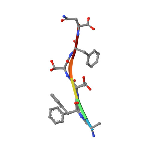
8DTS - PubMed Abstract:
J-domain protein (JDP) molecular chaperones have emerged as central players that maintain a healthy proteome. The diverse members of the JDP family function as monomers/dimers and a small subset assemble into micron-sized oligomers. The oligomeric JDP members have eluded structural characterization due to their low-complexity, intrinsically disordered middle domains. This in turn, obscures the biological significance of these larger oligomers in protein folding processes. Here, we identified a short, aromatic motif within DNAJB8 that drives self-assembly through π-π stacking and determined its X-ray structure. We show that mutations in the motif disrupt DNAJB8 oligomerization in vitro and in cells. DNAJB8 variants that are unable to assemble bind to misfolded tau seeds more specifically and retain capacity to reduce protein aggregation in vitro and in cells. We propose a new model for DNAJB8 function in which the sequences in the low-complexity domains play distinct roles in assembly and substrate activity.
- Center for Alzheimer's and Neurodegenerative Diseases, Peter O'Donnell Jr. Brain Institute, University of Texas Southwestern Medical Center, Dallas, TX 75390, USA.
Organizational Affiliation: