8DQJ | pdb_00008dqj

Crystal structure of pyrrolysyl-tRNA synthetase from Methanomethylophilus alvus engineered for acridone amino acid (AST) bound to ATP and acridone


Experimental Data Snapshot

  • Method: X-RAY DIFFRACTION
  • Resolution: 1.54 Å
  • R-Value Free: 
    0.193 (Depositor), 0.194 (DCC) 
  • R-Value Work: 
    0.170 (Depositor), 0.172 (DCC) 
  • R-Value Observed: 
    0.172 (Depositor) 

Starting Model: experimental
View more details

wwPDB Validation 3D Report Full Report

Validation slider image for 8DQJ

Ligand Structure Quality Assessment 


This is version 1.2 of the entry. See complete history

Literature

Structures of Methanomethylophilus alvus Pyrrolysine tRNA-Synthetases Support the Need for De Novo Selections When Altering the Substrate Specificity.

Gottfried-Lee, I.Perona, J.J.Karplus, P.A.Mehl, R.A.Cooley, R.B.

(2022) ACS Chem Biol 17: 3470-3477

  • DOI: https://doi.org/10.1021/acschembio.2c00640
  • Primary Citation Related Structures: 
    8DQG, 8DQH, 8DQI, 8DQJ

  • PubMed Abstract: 

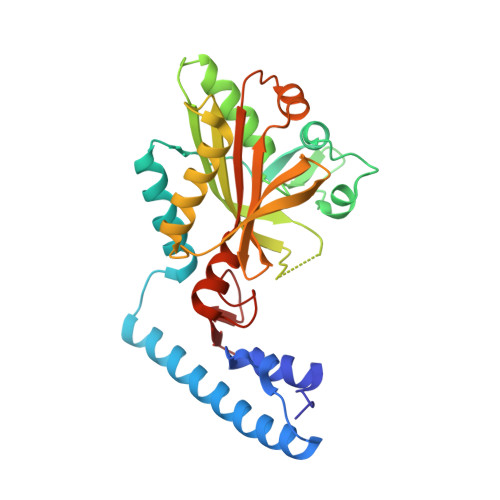
    A recently developed genetic code expansion (GCE) platform based on the pyrrolysine amino-acyl tRNA synthetase (PylRS)/tRNA Pyl pair from Methanomethylophilus alvus (Ma) has improved solubility and lower susceptibility to proteolysis compared with the homologous and commonly used Methanosarcina barkeri (Mb) and M. mazei (Mm) PylRS GCE platforms. We recently created two new Ma PylRS variants for the incorporation of the fluorescent amino acid, acridonyl-alanine (Acd), into proteins at amber codons: one based on "transplanting" active site mutations from an established high-efficiency Mb PylRS and one that was de novo selected from a library of mutants. Here, we present the crystal structures of these two Ma PylRS variants with Acd/ATP bound to understand why the "active site transplant" variant (Acd-AST) displayed 6-fold worse Acd incorporation efficiency than the de novo selected PylRS (called Acd-RS1). The structures reveal that the Acd-AST binding pocket is too small and binds the three-ring aromatic Acd in a distorted conformation, whereas the more spacious Acd-RS1 active site binds Acd in a relaxed, planar conformation stabilized by a network of solvent-mediated hydrogen bonds. The poor performance of the AST enzyme is ascribed to a shift in the Ma PylRS β-sheet framework relative to that of the Mb enzyme. This illustrates a general reason why "active site transplantation" may not succeed in creating efficient Ma PylRSs for other noncanonical amino acids. This work also provides structural details that will help guide the development of future Ma PylRS/tRNA Pyl GCE systems via de novo selection or directed evolution methods.


  • Organizational Affiliation
    • Department of Biochemistry and Biophysics, 2011 Agricultural and Life Sciences, Oregon State University, Corvallis, Oregon 97331, United States.

Macromolecule Content 

  • Total Structure Weight: 62.91 kDa 
  • Atom Count: 4,514 
  • Modeled Residue Count: 522 
  • Deposited Residue Count: 552 
  • Unique protein chains: 1

Macromolecules

Find similar proteins by:|  3D Structure
Entity ID: 1
MoleculeChains  Sequence LengthOrganismDetailsImage
AA_TRNA_LIGASE_II domain-containing proteinA [auth D],
B [auth A]
276Methanomethylophilus alviMutation(s): 3 
Gene Names: BKD89_05515
UniProt
Find proteins for A0A3G3IHP7 (Methanomethylophilus alvi)
Explore A0A3G3IHP7 
Go to UniProtKB:  A0A3G3IHP7
Entity Groups
Sequence Clusters30% Identity50% Identity70% Identity90% Identity95% Identity100% Identity
UniProt GroupA0A3G3IHP7
Sequence Annotations
Expand
Reference Sequence

Small Molecules

Ligands 6 Unique
IDChains Name / Formula / InChI Key2D Diagram3D Interactions
ATP

Query on ATP


Download:Ideal Coordinates CCD File
C [auth D]ADENOSINE-5'-TRIPHOSPHATE
C10 H16 N5 O13 P3
ZKHQWZAMYRWXGA-KQYNXXCUSA-N
AMP
(Subject of Investigation/LOI)

Query on AMP


Download:Ideal Coordinates CCD File
I [auth A]ADENOSINE MONOPHOSPHATE
C10 H14 N5 O7 P
UDMBCSSLTHHNCD-KQYNXXCUSA-N
T7Q
(Subject of Investigation/LOI)

Query on T7Q


Download:Ideal Coordinates CCD File
D
(2~{S})-2-azanyl-3-(9-oxidanylidene-10~{H}-acridin-2-yl)propanoic acid
C16 H15 N2 O3
NPALYBYZIAGBTE-LBPRGKRZSA-O
PEG

Query on PEG


Download:Ideal Coordinates CCD File
J [auth A]DI(HYDROXYETHYL)ETHER
C4 H10 O3
MTHSVFCYNBDYFN-UHFFFAOYSA-N
GOL

Query on GOL


Download:Ideal Coordinates CCD File
H [auth A]GLYCEROL
C3 H8 O3
PEDCQBHIVMGVHV-UHFFFAOYSA-N
MG

Query on MG


Download:Ideal Coordinates CCD File
E [auth D],
F [auth D],
G [auth D]
MAGNESIUM ION
Mg
JLVVSXFLKOJNIY-UHFFFAOYSA-N

Experimental Data & Validation

Experimental Data

  • Method: X-RAY DIFFRACTION
  • Resolution: 1.54 Å
  • R-Value Free:  0.193 (Depositor), 0.194 (DCC) 
  • R-Value Work:  0.170 (Depositor), 0.172 (DCC) 
  • R-Value Observed: 0.172 (Depositor) 
Space Group: I 4
Unit Cell:
Length ( Å )Angle ( ˚ )
a = 110.722α = 90
b = 110.722β = 90
c = 114.157γ = 90
Software Package:
Software NamePurpose
PHENIXrefinement
XDSdata reduction
Aimlessdata scaling
PHENIXphasing

Structure Validation

View Full Validation Report


Ligand Structure Quality Assessment 


Entry History 

& Funding Information

Deposition Data


Funding OrganizationLocationGrant Number
National Institutes of Health/National Institute of General Medical Sciences (NIH/NIGMS)United States1R01GM131168-01
National Institutes of Health/National Institute of General Medical Sciences (NIH/NIGMS)United StatesRM1-GM144227

Revision History  (Full details and data files)

  • Version 1.0: 2022-12-07
    Type: Initial release
  • Version 1.1: 2022-12-28
    Changes: Database references
  • Version 1.2: 2023-10-25
    Changes: Data collection, Refinement description