Crystal structures of NAD(P)H nitroreductases from Klebsiella pneumoniae.
Kancherla, A.D., Liu, L., Tillery, L., Shek, R., Craig, J.K., Machen, A.J., Seibold, S., Battaile, K.P., Fradi, S., Barrett, L.K., Subramanian, S., Myler, P., Van Voorhis, W.C., Lovell, S.(2024) Acta Crystallogr F Struct Biol Commun 80: 173-182
- PubMed: 38990055 Search on PubMedSearch on PubMed Central
- DOI: https://doi.org/10.1107/S2053230X24006472
- Primary Citation Related Structures:
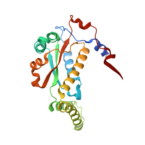
8DOR - PubMed Abstract:
Klebsiella pneumoniae (Kp) is an infectious disease pathogen that poses a significant global health threat due to its potential to cause severe infections and its tendency to exhibit multidrug resistance. Understanding the enzymatic mechanisms of the oxygen-insensitive nitroreductases (Kp-NRs) from Kp is crucial for the development of effective nitrofuran drugs, such as nitrofurantoin, that can be activated as antibiotics. In this paper, three crystal structures of two Kp-NRs (PDB entries 7tmf/7tmg and 8dor) are presented, and an analysis of their crystal structures and their flavin mononucleotide (FMN)-binding mode is provided. The structures with PDB codes 7tmf (Kp-NR1a), 7tmg (Kp-NR1b) and 8dor (Kp-NR2) were determined at resolutions of 1.97, 1.90 and 1.35 Å, respectively. The Kp-NR1a and Kp-NR1b structures adopt an αβ fold, in which four-stranded antiparallel β-sheets are surrounded by five helices. With domain swapping, the β-sheet was expanded with a β-strand from the other molecule of the dimer. The difference between the structures lies in the loop spanning Leu173-Ala185: in Kp-NR1a the loop is disordered, whereas the loop adopts multiple conformations in Kp-NR1b. The FMN interactions within Kp-NR1/NR2 involve hydrogen-bond and π-stacking interactions. Kp-NR2 contains four-stranded antiparallel β-sheets surrounded by eight helices with two short helices and one β-sheet. Structural and sequence alignments show that Kp-NR1a/b and Kp-NR2 are homologs of the Escherichia coli oxygen-insensitive NRs YdjA and NfnB and of Enterobacter cloacae NR, respectively. By homology inference from E. coli, Kp-NR1a/b and Kp-NR2 may detoxify polynitroaromatic compounds and Kp-NR2 may activate nitrofuran drugs to cause bactericidal activity through a ping-pong bi-bi mechanism, respectively.
- Division of Allergy and Infectious Diseases, Center for Emerging and Re-emerging Infectious Diseases, Department of Medicine, University of Washington, Seattle, WA 98109, USA.
Organizational Affiliation: