8DN4

Cryo-EM structure of human Glycine Receptor alpha-1 beta heteromer, glycine-bound state3(desensitized state)


Experimental Data Snapshot

  • Method: ELECTRON MICROSCOPY
  • Resolution: 4.10 Å
  • Aggregation State: PARTICLE 
  • Reconstruction Method: SINGLE PARTICLE 

wwPDB Validation   3D Report Full Report


This is version 1.2 of the entry. See complete history


Literature

Asymmetric gating of a human hetero-pentameric glycine receptor.

Liu, X.Wang, W.

(2023) Nat Commun 14: 6377-6377

  • DOI: https://doi.org/10.1038/s41467-023-42051-6
  • Primary Citation of Related Structures:  
    8DN2, 8DN3, 8DN4, 8DN5

  • PubMed Abstract: 

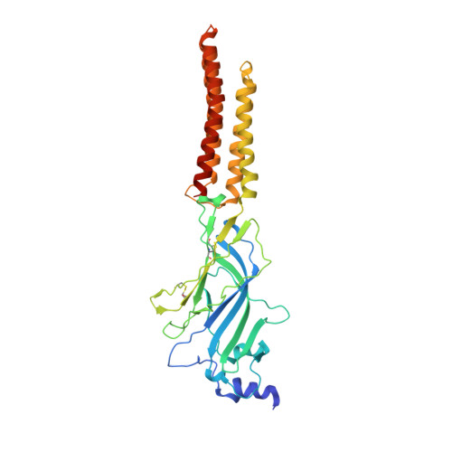
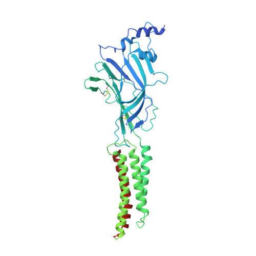
    Hetero-pentameric Cys-loop receptors constitute a major type of neurotransmitter receptors that enable signal transmission and processing in the nervous system. Despite intense investigations into their working mechanism and pharmaceutical potentials, how neurotransmitters activate these receptors remains unclear due to the lack of high-resolution structural information in the activated open state. Here we report near-atomic resolution structures resolved in digitonin consistent with all principle functional states of the human α1β GlyR, which is a major Cys-loop receptor that mediates inhibitory neurotransmission in the central nervous system of adults. Glycine binding induces cooperative and symmetric structural rearrangements in the neurotransmitter-binding extracellular domain but asymmetrical pore dilation in the transmembrane domain. Symmetric response in the extracellular domain is consistent with electrophysiological data showing cooperative glycine activation and contribution from both α1 and β subunits. A set of functionally essential but differentially charged amino acid residues in the transmembrane domain of the α1 and β subunits explains asymmetric activation. These findings provide a foundation for understanding how the gating of the Cys-loop receptor family members diverges to accommodate specific physiological environments.


  • Organizational Affiliation

    Department of Biophysics, University of Texas Southwestern Medical Center, Dallas, TX, USA.


Macromolecules
Find similar proteins by:  (by identity cutoff)  |  3D Structure
Entity ID: 1
MoleculeChains Sequence LengthOrganismDetailsImage
Glycine receptor subunit alpha-1A [auth D],
B [auth A],
C [auth B],
D [auth C]
367Homo sapiensMutation(s): 0 
Gene Names: GLRA1
Membrane Entity: Yes 
UniProt & NIH Common Fund Data Resources
Find proteins for P23415 (Homo sapiens)
Explore P23415 
Go to UniProtKB:  P23415
PHAROS:  P23415
GTEx:  ENSG00000145888 
Entity Groups  
Sequence Clusters30% Identity50% Identity70% Identity90% Identity95% Identity100% Identity
UniProt GroupP23415
Sequence Annotations
Expand
  • Reference Sequence
Find similar proteins by:  (by identity cutoff)  |  3D Structure
Entity ID: 2
MoleculeChains Sequence LengthOrganismDetailsImage
Glycine receptor subunit beta,Green fluorescent protein,Glycine receptor beta681Homo sapiensMutation(s): 0 
Gene Names: GLRBGFP
Membrane Entity: Yes 
UniProt & NIH Common Fund Data Resources
Find proteins for P42212 (Aequorea victoria)
Explore P42212 
Go to UniProtKB:  P42212
Find proteins for P48167 (Homo sapiens)
Explore P48167 
Go to UniProtKB:  P48167
PHAROS:  P48167
GTEx:  ENSG00000109738 
Entity Groups  
Sequence Clusters30% Identity50% Identity70% Identity90% Identity95% Identity100% Identity
UniProt GroupsP42212P48167
Sequence Annotations
Expand
  • Reference Sequence
Small Molecules
Ligands 6 Unique
IDChains Name / Formula / InChI Key2D Diagram3D Interactions
NAG
Query on NAG

Download Ideal Coordinates CCD File 
F [auth D],
K [auth A],
P [auth B],
S [auth C],
X [auth E]
2-acetamido-2-deoxy-beta-D-glucopyranose
C8 H15 N O6
OVRNDRQMDRJTHS-FMDGEEDCSA-N
DD9
Query on DD9

Download Ideal Coordinates CCD File 
T [auth C]nonane
C9 H20
BKIMMITUMNQMOS-UHFFFAOYSA-N
OCT
Query on OCT

Download Ideal Coordinates CCD File 
N [auth A]N-OCTANE
C8 H18
TVMXDCGIABBOFY-UHFFFAOYSA-N
HEX
Query on HEX

Download Ideal Coordinates CCD File 
H [auth D]
I [auth D]
L [auth A]
M [auth A]
O [auth A]
H [auth D],
I [auth D],
L [auth A],
M [auth A],
O [auth A],
Q [auth B],
R [auth B],
V [auth C],
Y [auth E]
HEXANE
C6 H14
VLKZOEOYAKHREP-UHFFFAOYSA-N
LNK
Query on LNK

Download Ideal Coordinates CCD File 
G [auth D]PENTANE
C5 H12
OFBQJSOFQDEBGM-UHFFFAOYSA-N
NBU
Query on NBU

Download Ideal Coordinates CCD File 
J [auth D],
U [auth C],
W [auth C]
N-BUTANE
C4 H10
IJDNQMDRQITEOD-UHFFFAOYSA-N
Experimental Data & Validation

Experimental Data

  • Method: ELECTRON MICROSCOPY
  • Resolution: 4.10 Å
  • Aggregation State: PARTICLE 
  • Reconstruction Method: SINGLE PARTICLE 
EM Software:
TaskSoftware PackageVersion
MODEL REFINEMENTPHENIX1.19.2_4158:

Structure Validation

View Full Validation Report


Entry History & Funding Information

Deposition Data


Funding OrganizationLocationGrant Number
National Institutes of Health/National Cancer Institute (NIH/NCI)United States1R35GM146860

Revision History  (Full details and data files)

  • Version 1.0: 2023-10-11
    Type: Initial release
  • Version 1.1: 2023-10-25
    Changes: Database references
  • Version 1.2: 2023-11-01
    Changes: Database references, Source and taxonomy, Structure summary