8DN1

Q108K:K40L:T51C:T53A:R58L:Q38F:Q4F mutant of hCRBPII bound to synthetic fluorophore CM1V at pH 7.2


Experimental Data Snapshot

  • Method: X-RAY DIFFRACTION
  • Resolution: 1.32 Å
  • R-Value Free: 0.217 
  • R-Value Work: 0.188 
  • R-Value Observed: 0.190 

wwPDB Validation   3D Report Full Report


Ligand Structure Quality Assessment 


This is version 1.3 of the entry. See complete history


Literature

Light controlled reversible Michael addition of cysteine: a new tool for dynamic site-specific labeling of proteins.

Maity, S.Bingham, C.Sheng, W.Ehyaei, N.Chakraborty, D.Tahmasebi-Nick, S.Kimmel, T.E.Vasileiou, C.Geiger, J.H.Borhan, B.

(2023) Analyst 148: 1085-1092

  • DOI: https://doi.org/10.1039/d2an01395a
  • Primary Citation of Related Structures:  
    8D6H, 8D6L, 8D6N, 8DB2, 8DN1

  • PubMed Abstract: 

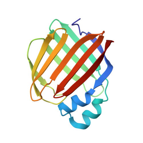
    Cysteine-based Michael addition is a widely employed strategy for covalent conjugation of proteins, peptides, and drugs. The covalent reaction is irreversible in most cases, leading to a lack of control over the process. Utilizing spectroscopic analyses along with X-ray crystallographic studies, we demonstrate Michael addition of an engineered cysteine residue in human Cellular Retinol Binding Protein II (hCRBPII) with a coumarin analog that creates a non-fluorescent complex. UV-illumination reverses the conjugation, yielding a fluorescent species, presumably through a retro -Michael process. This series of events can be repeated between a bound and non-bound form of the cysteine reversibly, resulting in the ON-OFF control of fluorescence. The details of the mechanism of photoswitching was illuminated by recapitulation of the process in light irradiated single crystals, confirming the mechanism at atomic resolution.


  • Organizational Affiliation

    Department of Chemistry, Michigan State University, 578 S. Shaw Ln., East Lansing, MI 48824, USA. babak@chemistry.msu.edu.


Macromolecules
Find similar proteins by:  (by identity cutoff)  |  3D Structure
Entity ID: 1
MoleculeChains Sequence LengthOrganismDetailsImage
Retinol-binding protein 2133Homo sapiensMutation(s): 7 
Gene Names: RBP2CRBP2
UniProt & NIH Common Fund Data Resources
Find proteins for P50120 (Homo sapiens)
Explore P50120 
Go to UniProtKB:  P50120
PHAROS:  P50120
GTEx:  ENSG00000114113 
Entity Groups  
Sequence Clusters30% Identity50% Identity70% Identity90% Identity95% Identity100% Identity
UniProt GroupP50120
Sequence Annotations
Expand
  • Reference Sequence
Experimental Data & Validation

Experimental Data

  • Method: X-RAY DIFFRACTION
  • Resolution: 1.32 Å
  • R-Value Free: 0.217 
  • R-Value Work: 0.188 
  • R-Value Observed: 0.190 
  • Space Group: C 1 2 1
Unit Cell:
Length ( Å )Angle ( ˚ )
a = 29.222α = 90
b = 66.833β = 91.926
c = 63.704γ = 90
Software Package:
Software NamePurpose
PHENIXrefinement
PHENIXrefinement
HKL-2000data reduction
HKL-2000data scaling
PHASERphasing

Structure Validation

View Full Validation Report


Ligand Structure Quality Assessment 


Entry History & Funding Information

Deposition Data


Funding OrganizationLocationGrant Number
National Institutes of Health/National Institute of Biomedical Imaging and Bioengineering (NIH/NIBIB)United States--

Revision History  (Full details and data files)

  • Version 1.0: 2023-02-01
    Type: Initial release
  • Version 1.1: 2023-02-15
    Changes: Database references
  • Version 1.2: 2023-03-15
    Changes: Database references
  • Version 1.3: 2023-10-25
    Changes: Data collection, Refinement description