Structural insights into actin isoforms.
Arora, A.S., Huang, H.L., Singh, R., Narui, Y., Suchenko, A., Hatano, T., Heissler, S.M., Balasubramanian, M.K., Chinthalapudi, K.(2023) Elife 12
- PubMed: 36790143 Search on PubMedSearch on PubMed Central
- DOI: https://doi.org/10.7554/eLife.82015
- Primary Citation Related Structures:
8DMX, 8DMY, 8DNF, 8DNH - PubMed Abstract:
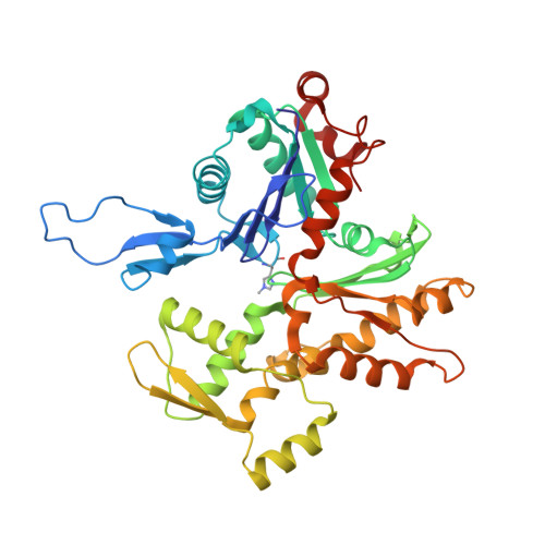
Actin isoforms organize into distinct networks that are essential for the normal function of eukaryotic cells. Despite a high level of sequence and structure conservation, subtle differences in their design principles determine the interaction with myosin motors and actin-binding proteins. Therefore, identifying how the structure of actin isoforms relates to function is important for our understanding of normal cytoskeletal physiology. Here, we report the high-resolution structures of filamentous skeletal muscle α-actin (3.37 Å), cardiac muscle α-actin (3.07 Å), ß-actin (2.99 Å), and γ-actin (3.38 Å) in the Mg 2+ ·ADP state with their native post-translational modifications. The structures revealed isoform-specific conformations of the N-terminus that shift closer to the filament surface upon myosin binding, thereby establishing isoform-specific interfaces. Collectively, the structures of single-isotype, post-translationally modified bare skeletal muscle α-actin, cardiac muscle α-actin, ß-actin, and γ-actin reveal general principles, similarities, and differences between isoforms. They complement the repertoire of known actin structures and allow for a comprehensive understanding of in vitro and in vivo functions of actin isoforms.
- Department of Physiology and Cell Biology, Dorothy M. Davis Heart and Lung Research Institute, The Ohio State University College of Medicine, Columbus, United States.
Organizational Affiliation: