8DL2 | pdb_00008dl2

BoGH13ASus from Bacteroides ovatus bound to acarbose


Experimental Data Snapshot

  • Method: X-RAY DIFFRACTION
  • Resolution: 1.99 Å
  • R-Value Free: 
    0.219 (Depositor), 0.223 (DCC) 
  • R-Value Work: 
    0.175 (Depositor), 0.184 (DCC) 
  • R-Value Observed: 
    0.178 (Depositor) 

Starting Model: experimental
View more details

wwPDB Validation 3D Report Full Report

Validation slider image for 8DL2

Ligand Structure Quality Assessment 


This is version 2.0 of the entry. See complete history

Literature

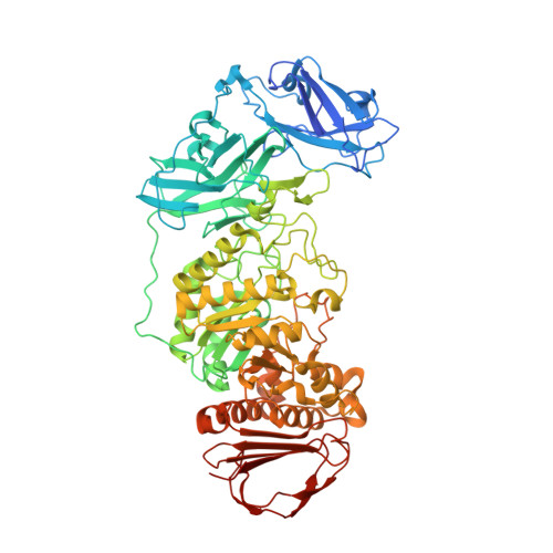
BoGH13A Sus from Bacteroides ovatus represents a novel alpha-amylase used for Bacteroides starch breakdown in the human gut.

Brown, H.A.DeVeaux, A.L.Juliano, B.R.Photenhauer, A.L.Boulinguiez, M.Bornschein, R.E.Wawrzak, Z.Ruotolo, B.T.Terrapon, N.Koropatkin, N.M.

(2023) Cell Mol Life Sci 80: 232-232

  • DOI: https://doi.org/10.1007/s00018-023-04812-w
  • Primary Citation Related Structures: 
    8DGE, 8DL1, 8DL2

  • PubMed Abstract: 

    Members of the Bacteroidetes phylum in the human colon deploy an extensive number of proteins to capture and degrade polysaccharides. Operons devoted to glycan breakdown and uptake are termed polysaccharide utilization loci or PUL. The starch utilization system (Sus) is one such PUL and was initially described in Bacteroides thetaiotaomicron (Bt). BtSus is highly conserved across many species, except for its extracellular α-amylase, SusG. In this work, we show that the Bacteroides ovatus (Bo) extracellular α-amylase, BoGH13A Sus , is distinguished from SusG in its evolutionary origin and its domain architecture and by being the most prevalent form in Bacteroidetes Sus. BoGH13A Sus is the founding member of both a novel subfamily in the glycoside hydrolase family 13, GH13_47, and a novel carbohydrate-binding module, CBM98. The BoGH13A Sus CBM98-CBM48-GH13_47 architecture differs from the CBM58 embedded within the GH13_36 of SusG. These domains adopt a distinct spatial orientation and invoke a different association with the outer membrane. The BoCBM98 binding site is required for Bo growth on polysaccharides and optimal enzymatic degradation thereof. Finally, the BoGH13A Sus structure features bound Ca 2+ and Mn 2+ ions, the latter of which is novel for an α-amylase. Little is known about the impact of Mn 2+ on gut bacterial function, much less on polysaccharide consumption, but Mn 2+ addition to Bt expressing BoGH13A Sus specifically enhances growth on starch. Further understanding of bacterial starch degradation signatures will enable more tailored prebiotic and pharmaceutical approaches that increase starch flux to the gut.


  • Organizational Affiliation
    • Department of Microbiology and Immunology, University of Michigan Medical School, Ann Arbor, MI, 48109, USA. haleybr@umich.edu.

Macromolecule Content 

  • Total Structure Weight: 346.94 kDa 
  • Atom Count: 25,774 
  • Modeled Residue Count: 2,845 
  • Deposited Residue Count: 2,952 
  • Unique protein chains: 1

Macromolecules

Find similar proteins by:|  3D Structure
Entity ID: 1
MoleculeChains  Sequence LengthOrganismDetailsImage
Alpha amylase, catalytic domain protein
A, B, C, D
738Bacteroides ovatus ATCC 8483Mutation(s): 0 
Gene Names: BACOVA_03514Bovatus_03803

Oligosaccharides

Help  
Entity ID: 2
MoleculeChains Length2D Diagram GlycosylationD Interactions
4,6-dideoxy-4-{[(1S,4R,5S,6S)-4,5,6-trihydroxy-3-(hydroxymethyl)cyclohex-2-en-1-yl]amino}-alpha-D-glucopyranose-(1-4)-alpha-D-glucopyranose-(1-4)-beta-D-glucopyranoseE [auth H],
H [auth K]
3N/AN/A
Entity ID: 3
MoleculeChains Length2D Diagram GlycosylationD Interactions
4,6-dideoxy-4-{[(1S,4R,5S,6S)-4,5,6-trihydroxy-3-(hydroxymethyl)cyclohex-2-en-1-yl]amino}-alpha-D-glucopyranose-(1-4)-alpha-D-glucopyranose-(1-4)-alpha-D-glucopyranose
F [auth I],
I [auth L],
J [auth O],
N,
O [auth V],
F [auth I],
I [auth L],
J [auth O],
N,
O [auth V],
P [auth Q]
3N/AN/A
Entity ID: 4
MoleculeChains Length2D Diagram GlycosylationD Interactions
alpha-D-glucopyranose-(1-4)-alpha-D-glucopyranose-(1-4)-4,6-dideoxy-4-{[(1S,4R,5S,6S)-4,5,6-trihydroxy-3-(hydroxymethyl)cyclohex-2-en-1-yl]amino}-alpha-D-glucopyranose-(1-4)-alpha-D-glucopyranose-(1-4)-alpha-D-glucopyranoseG [auth J]5N/AN/A
Entity ID: 5
MoleculeChains Length2D Diagram GlycosylationD Interactions
alpha-D-glucopyranose-(1-4)-alpha-D-glucopyranoseK [auth P]2N/AN/A
Entity ID: 6
MoleculeChains Length2D Diagram GlycosylationD Interactions
4,6-dideoxy-4-{[(1S,4R,5S,6S)-4,5,6-trihydroxy-3-(hydroxymethyl)cyclohex-2-en-1-yl]amino}-alpha-D-glucopyranose-(1-4)-alpha-D-glucopyranoseL [auth R]2N/AN/A
Entity ID: 7
MoleculeChains Length2D Diagram GlycosylationD Interactions
alpha-D-glucopyranose-(1-4)-4,6-dideoxy-4-{[(1S,4R,5S,6S)-4,5,6-trihydroxy-3-(hydroxymethyl)cyclohex-2-en-1-yl]amino}-alpha-D-glucopyranose-(1-4)-alpha-D-glucopyranose-(1-4)-alpha-D-glucopyranoseM,
Q [auth S]
4N/AN/A

Small Molecules

Ligands 9 Unique
IDChains Name / Formula / InChI Key2D Diagram3D Interactions
GLC
(Subject of Investigation/LOI)

Query on GLC


Download:Ideal Coordinates CCD File
R [auth A],
Y [auth B]
alpha-D-glucopyranose
C6 H12 O6
WQZGKKKJIJFFOK-DVKNGEFBSA-N
PGE

Query on PGE


Download:Ideal Coordinates CCD File
EA [auth B],
T [auth A]
TRIETHYLENE GLYCOL
C6 H14 O4
ZIBGPFATKBEMQZ-UHFFFAOYSA-N
PEG

Query on PEG


Download:Ideal Coordinates CCD File
BA [auth B],
OA [auth D]
DI(HYDROXYETHYL)ETHER
C4 H10 O3
MTHSVFCYNBDYFN-UHFFFAOYSA-N
GOL

Query on GOL


Download:Ideal Coordinates CCD File
AA [auth B]
CA [auth B]
DA [auth B]
IA [auth C]
JA [auth C]
AA [auth B],
CA [auth B],
DA [auth B],
IA [auth C],
JA [auth C],
U [auth A]
GLYCEROL
C3 H8 O3
PEDCQBHIVMGVHV-UHFFFAOYSA-N
EDO

Query on EDO


Download:Ideal Coordinates CCD File
PA [auth D],
V [auth A]
1,2-ETHANEDIOL
C2 H6 O2
LYCAIKOWRPUZTN-UHFFFAOYSA-N
ACT

Query on ACT


Download:Ideal Coordinates CCD File
FA [auth B],
KA [auth C],
W [auth A]
ACETATE ION
C2 H3 O2
QTBSBXVTEAMEQO-UHFFFAOYSA-M
MN

Query on MN


Download:Ideal Coordinates CCD File
GA [auth B],
LA [auth C],
QA [auth D],
X [auth A]
MANGANESE (II) ION
Mn
WAEMQWOKJMHJLA-UHFFFAOYSA-N
CA

Query on CA


Download:Ideal Coordinates CCD File
HA [auth C],
MA [auth D],
S [auth A],
Z [auth B]
CALCIUM ION
Ca
BHPQYMZQTOCNFJ-UHFFFAOYSA-N
CL

Query on CL


Download:Ideal Coordinates CCD File
NA [auth D]CHLORIDE ION
Cl
VEXZGXHMUGYJMC-UHFFFAOYSA-M

Experimental Data & Validation

Experimental Data

  • Method: X-RAY DIFFRACTION
  • Resolution: 1.99 Å
  • R-Value Free:  0.219 (Depositor), 0.223 (DCC) 
  • R-Value Work:  0.175 (Depositor), 0.184 (DCC) 
  • R-Value Observed: 0.178 (Depositor) 
Space Group: P 1 21 1
Unit Cell:
Length ( Å )Angle ( ˚ )
a = 100.08α = 90
b = 125.31β = 102.11
c = 150.78γ = 90
Software Package:
Software NamePurpose
REFMACrefinement
Aimlessdata scaling
PDB_EXTRACTdata extraction
xia2data reduction
PHASERphasing

Structure Validation

View Full Validation Report


Ligand Structure Quality Assessment 


Entry History 

& Funding Information

Deposition Data


Funding OrganizationLocationGrant Number
National Institutes of Health/National Institute of General Medical Sciences (NIH/NIGMS)United StatesGM118475
National Institutes of Health/National Center for Complementary and Integrative Health (NIH/NCCIH)United StatesAT011278

Revision History  (Full details and data files)

  • Version 1.0: 2023-05-24
    Type: Initial release
  • Version 1.1: 2023-09-20
    Changes: Data collection, Database references
  • Version 1.2: 2023-10-25
    Changes: Refinement description
  • Version 2.0: 2024-04-10
    Changes: Atomic model, Derived calculations