


Sulfide oxidation in human sulfide quinone oxidoreductase is enthalpically driven: Contributions of the Lys-207 general base
Bonanata, J., Landry, A., Ceric, K., Ludlam, A., Mascarenhas, R., Coitino, L., Banerjee, R.To be published.
Experimental Data Snapshot
Starting Model: experimental
View more details
Entity ID: 1 | |||||
|---|---|---|---|---|---|
| Molecule | Chains | Sequence Length | Organism | Details | Image |
| Sulfide:quinone oxidoreductase | 418 | Homo sapiens | Mutation(s): 1 EC: 1.8.5.8 | 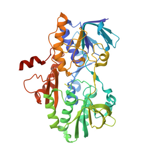 | |
UniProt & NIH Common Fund Data Resources | |||||
PHAROS: Q9Y6N5 GTEx: ENSG00000137767 | |||||
Entity Groups | |||||
| Sequence Clusters | 30% Identity50% Identity70% Identity90% Identity95% Identity100% Identity | ||||
| UniProt Group | Q9Y6N5 | ||||
Sequence AnnotationsExpand | |||||
Reference Sequence | |||||
| Ligands 4 Unique | |||||
|---|---|---|---|---|---|
| ID | Chains | Name / Formula / InChI Key | 2D Diagram | 3D Interactions | |
| FAD (Subject of Investigation/LOI) Download:Ideal Coordinates CCD File | C [auth A], K [auth B] | FLAVIN-ADENINE DINUCLEOTIDE C27 H33 N9 O15 P2 VWWQXMAJTJZDQX-UYBVJOGSSA-N |  | ||
| MHA Download:Ideal Coordinates CCD File | E [auth A], Q [auth B] | (CARBAMOYLMETHYL-CARBOXYMETHYL-AMINO)-ACETIC ACID C6 H10 N2 O5 QZTKDVCDBIDYMD-UHFFFAOYSA-N |  | ||
| PEG Download:Ideal Coordinates CCD File | D [auth A] F [auth A] G [auth A] I [auth A] J [auth A] | DI(HYDROXYETHYL)ETHER C4 H10 O3 MTHSVFCYNBDYFN-UHFFFAOYSA-N |  | ||
| SO4 Download:Ideal Coordinates CCD File | H [auth A], R [auth B], S [auth B], T [auth B] | SULFATE ION O4 S QAOWNCQODCNURD-UHFFFAOYSA-L |  | ||
| Modified Residues 1 Unique | |||||
|---|---|---|---|---|---|
| ID | Chains | Type | Formula | 2D Diagram | Parent |
| CSS Query on CSS | A, B | L-PEPTIDE LINKING | C3 H7 N O2 S2 |  | CYS |
| Length ( Å ) | Angle ( ˚ ) |
|---|---|
| a = 76.793 | α = 90 |
| b = 111.298 | β = 90 |
| c = 134.105 | γ = 90 |
| Software Name | Purpose |
|---|---|
| PHENIX | refinement |
| Aimless | data scaling |
| PDB_EXTRACT | data extraction |
| DIALS | data reduction |
| PHASER | phasing |
| Funding Organization | Location | Grant Number |
|---|---|---|
| National Institutes of Health/National Institute of General Medical Sciences (NIH/NIGMS) | United States | 5R35GM130183 |


