8DH9

Leptin-bound leptin receptor complex-D3-D7


Experimental Data Snapshot

  • Method: ELECTRON MICROSCOPY
  • Resolution: 4.50 Å
  • Aggregation State: PARTICLE 
  • Reconstruction Method: SINGLE PARTICLE 

wwPDB Validation   3D Report Full Report


This is version 1.0 of the entry. See complete history


Literature

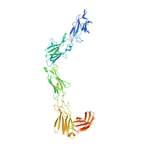
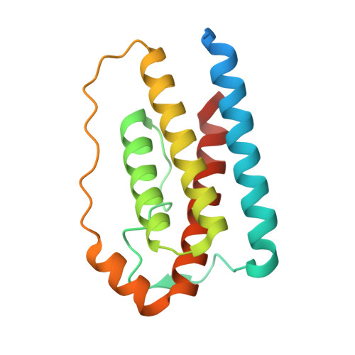
Structural insights into the mechanism of leptin receptor activation.

Saxton, R.A.Caveney, N.A.Moya-Garzon, M.D.Householder, K.D.Rodriguez, G.E.Burdsall, K.A.Long, J.Z.Garcia, K.C.

(2023) Nat Commun 14: 1797-1797

  • DOI: https://doi.org/10.1038/s41467-023-37169-6
  • Primary Citation of Related Structures:  
    8DH8, 8DH9, 8DHA

  • PubMed Abstract: 

    Leptin is an adipocyte-derived protein hormone that promotes satiety and energy homeostasis by activating the leptin receptor (LepR)-STAT3 signaling axis in a subset of hypothalamic neurons. Leptin signaling is dysregulated in obesity, however, where appetite remains elevated despite high levels of circulating leptin. To gain insight into the mechanism of leptin receptor activation, here we determine the structure of a stabilized leptin-bound LepR signaling complex using single particle cryo-EM. The structure reveals an asymmetric architecture in which a single leptin induces LepR dimerization via two distinct receptor-binding sites. Analysis of the leptin-LepR binding interfaces reveals the molecular basis for human obesity-associated mutations. Structure-based design of leptin variants that destabilize the asymmetric LepR dimer yield both partial and biased agonists that partially suppress STAT3 activation in the presence of wild-type leptin and decouple activation of STAT3 from LepR negative regulators. Together, these results reveal the structural basis for LepR activation and provide insights into the differential plasticity of signaling pathways downstream of LepR.


  • Organizational Affiliation

    Department of Molecular and Cellular Physiology, Stanford University School of Medicine, Stanford, CA, 94305, USA. rsaxton@berkeley.edu.


Macromolecules
Find similar proteins by:  (by identity cutoff)  |  3D Structure
Entity ID: 1
MoleculeChains Sequence LengthOrganismDetailsImage
Leptin receptor
A, B
557Mus musculusMutation(s): 0 
Gene Names: LeprDbObr
Membrane Entity: Yes 
UniProt & NIH Common Fund Data Resources
Find proteins for P48356 (Mus musculus)
Explore P48356 
Go to UniProtKB:  P48356
IMPC:  MGI:104993
Entity Groups  
Sequence Clusters30% Identity50% Identity70% Identity90% Identity95% Identity100% Identity
UniProt GroupP48356
Sequence Annotations
Expand
  • Reference Sequence
Find similar proteins by:  (by identity cutoff)  |  3D Structure
Entity ID: 2
MoleculeChains Sequence LengthOrganismDetailsImage
Leptin
C, D
167Mus musculusMutation(s): 0 
Gene Names: LepOb
Membrane Entity: Yes 
UniProt & NIH Common Fund Data Resources
Find proteins for P41160 (Mus musculus)
Explore P41160 
Go to UniProtKB:  P41160
IMPC:  MGI:104663
Entity Groups  
Sequence Clusters30% Identity50% Identity70% Identity90% Identity95% Identity100% Identity
UniProt GroupP41160
Sequence Annotations
Expand
  • Reference Sequence
Experimental Data & Validation

Experimental Data

  • Method: ELECTRON MICROSCOPY
  • Resolution: 4.50 Å
  • Aggregation State: PARTICLE 
  • Reconstruction Method: SINGLE PARTICLE 

Structure Validation

View Full Validation Report


Entry History & Funding Information

Deposition Data


Funding OrganizationLocationGrant Number
National Institutes of Health/National Institute Of Allergy and Infectious Diseases (NIH/NIAID)United States--

Revision History  (Full details and data files)

  • Version 1.0: 2023-04-19
    Type: Initial release