8DGP

14-3-3 epsilon bound to phosphorylated PEAK3 (pS69) peptide


Experimental Data Snapshot

  • Method: X-RAY DIFFRACTION
  • Resolution: 2.70 Å
  • R-Value Free: 0.233 
  • R-Value Work: 0.187 
  • R-Value Observed: 0.190 

wwPDB Validation   3D Report Full Report


This is version 1.2 of the entry. See complete history


Literature

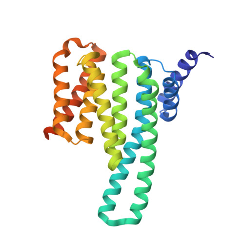
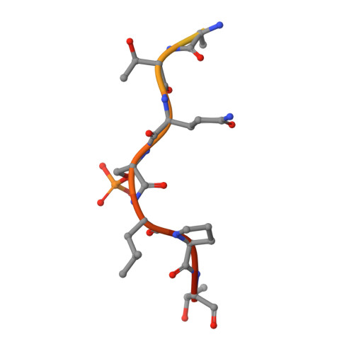
Structural mapping of PEAK pseudokinase interactions identifies 14-3-3 as a molecular switch for PEAK3 signaling.

Roy, M.J.Surudoi, M.G.Kropp, A.Hou, J.Dai, W.Hardy, J.M.Liang, L.Y.Cotton, T.R.Lechtenberg, B.C.Dite, T.A.Ma, X.Daly, R.J.Patel, O.Lucet, I.S.

(2023) Nat Commun 14: 3542-3542

  • DOI: https://doi.org/10.1038/s41467-023-38869-9
  • Primary Citation of Related Structures:  
    8DGM, 8DGN, 8DGO, 8DGP

  • PubMed Abstract: 

    PEAK pseudokinases regulate cell migration, invasion and proliferation by recruiting key signaling proteins to the cytoskeleton. Despite lacking catalytic activity, alteration in their expression level is associated with several aggressive cancers. Here, we elucidate the molecular details of key PEAK signaling interactions with the adapter proteins CrkII and Grb2 and the scaffold protein 14-3-3. Our findings rationalize why the dimerization of PEAK proteins has a crucial function in signal transduction and provide biophysical and structural data to unravel binding specificity within the PEAK interactome. We identify a conserved high affinity 14-3-3 motif on PEAK3 and demonstrate its role as a molecular switch to regulate CrkII binding and signaling via Grb2. Together, our studies provide a detailed structural snapshot of PEAK interaction networks and further elucidate how PEAK proteins, especially PEAK3, act as dynamic scaffolds that exploit adapter proteins to control signal transduction in cell growth/motility and cancer.


  • Organizational Affiliation

    The Walter and Eliza Hall Institute of Medical Research, Parkville, VIC, 3052, Australia. roy@wehi.edu.au.


Macromolecules
Find similar proteins by:  (by identity cutoff)  |  3D Structure
Entity ID: 1
MoleculeChains Sequence LengthOrganismDetailsImage
14-3-3 protein epsilon
A, B, C, D
258Homo sapiensMutation(s): 0 
Gene Names: YWHAE
UniProt & NIH Common Fund Data Resources
Find proteins for P62258 (Homo sapiens)
Explore P62258 
Go to UniProtKB:  P62258
PHAROS:  P62258
GTEx:  ENSG00000108953 
Entity Groups  
Sequence Clusters30% Identity50% Identity70% Identity90% Identity95% Identity100% Identity
UniProt GroupP62258
Sequence Annotations
Expand
  • Reference Sequence

Find similar proteins by:  Sequence   |   3D Structure  

Entity ID: 2
MoleculeChains Sequence LengthOrganismDetailsImage
Phosphorylated PEAK3 (pS69) peptide
E, F, G, H
21Homo sapiensMutation(s): 0 
UniProt & NIH Common Fund Data Resources
Find proteins for Q6ZS72 (Homo sapiens)
Explore Q6ZS72 
Go to UniProtKB:  Q6ZS72
GTEx:  ENSG00000188305 
Entity Groups  
Sequence Clusters30% Identity50% Identity70% Identity90% Identity95% Identity100% Identity
UniProt GroupQ6ZS72
Sequence Annotations
Expand
  • Reference Sequence
Experimental Data & Validation

Experimental Data

  • Method: X-RAY DIFFRACTION
  • Resolution: 2.70 Å
  • R-Value Free: 0.233 
  • R-Value Work: 0.187 
  • R-Value Observed: 0.190 
  • Space Group: P 41
Unit Cell:
Length ( Å )Angle ( ˚ )
a = 155.532α = 90
b = 155.532β = 90
c = 58.06γ = 90
Software Package:
Software NamePurpose
PHENIXrefinement
XDSdata reduction
Aimlessdata scaling
PHASERphasing
PDB_EXTRACTdata extraction
Cootmodel building

Structure Validation

View Full Validation Report


Entry History & Funding Information

Deposition Data


Funding OrganizationLocationGrant Number
National Health and Medical Research Council (NHMRC, Australia)AustraliaAPP1144149

Revision History  (Full details and data files)

  • Version 1.0: 2023-06-07
    Type: Initial release
  • Version 1.1: 2023-07-12
    Changes: Data collection, Database references
  • Version 1.2: 2023-10-25
    Changes: Data collection, Refinement description