8DBA

Crystal structure of dodecameric KaiC


Experimental Data Snapshot

  • Method: X-RAY DIFFRACTION
  • Resolution: 3.50 Å
  • R-Value Free: 0.279 
  • R-Value Work: 0.236 
  • R-Value Observed: 0.236 

wwPDB Validation   3D Report Full Report


Ligand Structure Quality Assessment 


This is version 1.3 of the entry. See complete history


Literature

From primordial clocks to circadian oscillators.

Pitsawong, W.Padua, R.A.P.Grant, T.Hoemberger, M.Otten, R.Bradshaw, N.Grigorieff, N.Kern, D.

(2023) Nature 616: 183-189

  • DOI: https://doi.org/10.1038/s41586-023-05836-9
  • Primary Citation of Related Structures:  
    8DB3, 8DBA, 8FWI, 8FWJ

  • PubMed Abstract: 

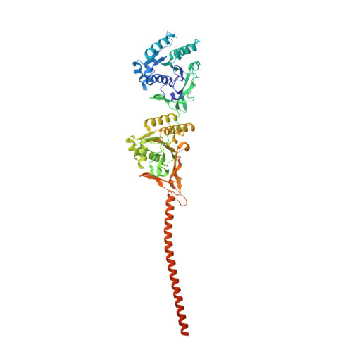
    Circadian rhythms play an essential part in many biological processes, and only three prokaryotic proteins are required to constitute a true post-translational circadian oscillator 1 . The evolutionary history of the three Kai proteins indicates that KaiC is the oldest member and a central component of the clock 2 . Subsequent additions of KaiB and KaiA regulate the phosphorylation state of KaiC for time synchronization. The canonical KaiABC system in cyanobacteria is well understood 3-6 , but little is known about more ancient systems that only possess KaiBC. However, there are reports that they might exhibit a basic, hourglass-like timekeeping mechanism 7-9 . Here we investigate the primordial circadian clock in Rhodobacter sphaeroides, which contains only KaiBC, to elucidate its inner workings despite missing KaiA. Using a combination of X-ray crystallography and cryogenic electron microscopy, we find a new dodecameric fold for KaiC, in which two hexamers are held together by a coiled-coil bundle of 12 helices. This interaction is formed by the carboxy-terminal extension of KaiC and serves as an ancient regulatory moiety that is later superseded by KaiA. A coiled-coil register shift between daytime and night-time conformations is connected to phosphorylation sites through a long-range allosteric network that spans over 140 Å. Our kinetic data identify the difference in the ATP-to-ADP ratio between day and night as the environmental cue that drives the clock. They also unravel mechanistic details that shed light on the evolution of self-sustained oscillators.


  • Organizational Affiliation

    Howard Hughes Medical Institute and Department of Biochemistry, Brandeis University, Waltham, MA, USA.


Macromolecules
Find similar proteins by:  (by identity cutoff)  |  3D Structure
Entity ID: 1
MoleculeChains Sequence LengthOrganismDetailsImage
Circadian clock protein KaiC
A, B, C, D, E
A, B, C, D, E, F, G, H, I, J, K, L
568Cereibacter sphaeroidesMutation(s): 0 
UniProt
Find proteins for B9KWX8 (Cereibacter sphaeroides (strain KD131 / KCTC 12085))
Explore B9KWX8 
Go to UniProtKB:  B9KWX8
Entity Groups  
Sequence Clusters30% Identity50% Identity70% Identity90% Identity95% Identity100% Identity
UniProt GroupB9KWX8
Sequence Annotations
Expand
  • Reference Sequence
Small Molecules
Ligands 2 Unique
IDChains Name / Formula / InChI Key2D Diagram3D Interactions
ADP (Subject of Investigation/LOI)
Query on ADP

Download Ideal Coordinates CCD File 
AB [auth J]
BB [auth K]
CA [auth D]
DA [auth E]
EB [auth K]
AB [auth J],
BB [auth K],
CA [auth D],
DA [auth E],
EB [auth K],
FB [auth L],
GA [auth E],
GB [auth L],
HA [auth F],
JA [auth F],
KA [auth G],
M [auth A],
NA [auth G],
P [auth A],
PA [auth H],
Q [auth B],
RA [auth H],
T [auth B],
TA [auth I],
V [auth C],
WA [auth I],
XA [auth J],
Y [auth C],
Z [auth D]
ADENOSINE-5'-DIPHOSPHATE
C10 H15 N5 O10 P2
XTWYTFMLZFPYCI-KQYNXXCUSA-N
MG (Subject of Investigation/LOI)
Query on MG

Download Ideal Coordinates CCD File 
AA [auth D]
BA [auth D]
CB [auth K]
DB [auth K]
EA [auth E]
AA [auth D],
BA [auth D],
CB [auth K],
DB [auth K],
EA [auth E],
FA [auth E],
IA [auth F],
LA [auth G],
MA [auth G],
N [auth A],
O [auth A],
OA [auth G],
QA [auth H],
R [auth B],
S [auth B],
SA [auth H],
U [auth B],
UA [auth I],
VA [auth I],
W [auth C],
X [auth C],
YA [auth J],
ZA [auth J]
MAGNESIUM ION
Mg
JLVVSXFLKOJNIY-UHFFFAOYSA-N
Experimental Data & Validation

Experimental Data

  • Method: X-RAY DIFFRACTION
  • Resolution: 3.50 Å
  • R-Value Free: 0.279 
  • R-Value Work: 0.236 
  • R-Value Observed: 0.236 
  • Space Group: P 1
Unit Cell:
Length ( Å )Angle ( ˚ )
a = 105.11α = 93.13
b = 136.09β = 94.43
c = 146.2γ = 108.09
Software Package:
Software NamePurpose
PHENIXrefinement
MOSFLMdata reduction
Aimlessdata scaling
PHASERphasing

Structure Validation

View Full Validation Report


Ligand Structure Quality Assessment 


Entry History & Funding Information

Deposition Data


Funding OrganizationLocationGrant Number
Howard Hughes Medical Institute (HHMI)United States--

Revision History  (Full details and data files)

  • Version 1.0: 2023-03-22
    Type: Initial release
  • Version 1.1: 2023-04-05
    Changes: Database references
  • Version 1.2: 2023-04-19
    Changes: Database references
  • Version 1.3: 2023-10-25
    Changes: Data collection, Database references, Refinement description