8D6M

Nanorana parkeri saxiphilin:STX (co-crystal)


Experimental Data Snapshot

  • Method: X-RAY DIFFRACTION
  • Resolution: 2.00 Å
  • R-Value Free: 0.220 
  • R-Value Work: 0.188 
  • R-Value Observed: 0.190 

wwPDB Validation   3D Report Full Report


Ligand Structure Quality Assessment 


This is version 2.0 of the entry. See complete history


Literature

Definition of a saxitoxin (STX) binding code enables discovery and characterization of the anuran saxiphilin family.

Chen, Z.Zakrzewska, S.Hajare, H.S.Alvarez-Buylla, A.Abderemane-Ali, F.Bogan, M.Ramirez, D.O'Connell, L.A.Du Bois, J.Minor Jr., D.L.

(2022) Proc Natl Acad Sci U S A 119: e2210114119-e2210114119

  • DOI: https://doi.org/10.1073/pnas.2210114119
  • Primary Citation of Related Structures:  
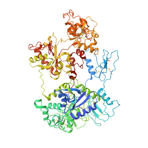
    8D6G, 8D6M, 8D6O, 8D6P, 8D6Q, 8D6S, 8D6T, 8D6U

  • PubMed Abstract: 

    American bullfrog ( Rana castesbeiana ) saxiphilin ( Rc Sxph) is a high-affinity "toxin sponge" protein thought to prevent intoxication by saxitoxin (STX), a lethal bis-guanidinium neurotoxin that causes paralytic shellfish poisoning (PSP) by blocking voltage-gated sodium channels (Na V s). How specific Rc Sxph interactions contribute to STX binding has not been defined and whether other organisms have similar proteins is unclear. Here, we use mutagenesis, ligand binding, and structural studies to define the energetic basis of Sxph:STX recognition. The resultant STX "recognition code" enabled engineering of Rc Sxph to improve its ability to rescue Na V s from STX and facilitated discovery of 10 new frog and toad Sxphs. Definition of the STX binding code and Sxph family expansion among diverse anurans separated by ∼140 My of evolution provides a molecular basis for understanding the roles of toxin sponge proteins in toxin resistance and for developing novel proteins to sense or neutralize STX and related PSP toxins.


  • Organizational Affiliation

    Cardiovascular Research Institute, University of California, San Francisco, CA 94158.


Macromolecules
Find similar proteins by:  (by identity cutoff)  |  3D Structure
Entity ID: 1
MoleculeChains Sequence LengthOrganismDetailsImage
Saxiphilin854Nanorana parkeriMutation(s): 0 
Gene Names: XM_018555331.1
Entity Groups  
Sequence Clusters30% Identity50% Identity70% Identity90% Identity95% Identity100% Identity
Sequence Annotations
Expand
  • Reference Sequence
Small Molecules
Ligands 2 Unique
IDChains Name / Formula / InChI Key2D Diagram3D Interactions
9SL (Subject of Investigation/LOI)
Query on 9SL

Download Ideal Coordinates CCD File 
C [auth A][(3aS,4R,10aS)-2,6-diamino-10,10-dihydroxy-3a,4,9,10-tetrahydro-3H,8H-pyrrolo[1,2-c]purin-4-yl]methyl carbamate
C10 H17 N7 O4
RPQXVSUAYFXFJA-HGRQIUPRSA-N
1PE
Query on 1PE

Download Ideal Coordinates CCD File 
B [auth A]PENTAETHYLENE GLYCOL
C10 H22 O6
JLFNLZLINWHATN-UHFFFAOYSA-N
Experimental Data & Validation

Experimental Data

  • Method: X-RAY DIFFRACTION
  • Resolution: 2.00 Å
  • R-Value Free: 0.220 
  • R-Value Work: 0.188 
  • R-Value Observed: 0.190 
  • Space Group: H 3
Unit Cell:
Length ( Å )Angle ( ˚ )
a = 228.848α = 90
b = 228.848β = 90
c = 67.224γ = 120
Software Package:
Software NamePurpose
PHENIXrefinement
Aimlessdata scaling
PDB_EXTRACTdata extraction
XDSdata reduction
PHASERphasing

Structure Validation

View Full Validation Report


Ligand Structure Quality Assessment 


Entry History & Funding Information

Deposition Data


Funding OrganizationLocationGrant Number
Department of Defense (DOD, United States)United StatesHDTRA-1-19-1-0040
Department of Defense (DOD, United States)United StatesHDTRA-1-21-1-10011

Revision History  (Full details and data files)

  • Version 1.0: 2022-11-02
    Type: Initial release
  • Version 1.1: 2023-10-18
    Changes: Data collection, Refinement description
  • Version 2.0: 2024-03-27
    Type: Coordinate replacement
    Reason: Ligand geometry
    Changes: Advisory, Atomic model, Data collection, Derived calculations, Refinement description, Structure summary