


Crystal structure of dihydropteroate synthase (folP-SMZ_B27) from soil uncultured bacterium in complex with 6-hydroxymethyl-7,8-dihydropterin
Stogios, P.J.To be published.
Experimental Data Snapshot
Starting Model: experimental
View more details
Entity ID: 1 | |||||
|---|---|---|---|---|---|
| Molecule | Chains | Sequence Length | Organism | Details | Image |
| folP-SMZ_B27 | 274 | uncultured bacterium | Mutation(s): 0 Gene Names: folP-SMZ_B27 | 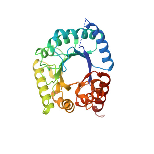 | |
| Ligands 4 Unique | |||||
|---|---|---|---|---|---|
| ID | Chains | Name / Formula / InChI Key | 2D Diagram | 3D Interactions | |
| HHR (Subject of Investigation/LOI) Download:Ideal Coordinates CCD File | E [auth A], M [auth B] | 6-HYDROXYMETHYLPTERIN C7 H7 N5 O2 XGWIBNWDLMIPNF-UHFFFAOYSA-N |  | ||
| PEG Download:Ideal Coordinates CCD File | C [auth A], D [auth A], J [auth B], K [auth B], L [auth B] | DI(HYDROXYETHYL)ETHER C4 H10 O3 MTHSVFCYNBDYFN-UHFFFAOYSA-N |  | ||
| SO4 Download:Ideal Coordinates CCD File | F [auth A], G [auth A], H [auth A], N [auth B] | SULFATE ION O4 S QAOWNCQODCNURD-UHFFFAOYSA-L |  | ||
| CL Download:Ideal Coordinates CCD File | I [auth A] | CHLORIDE ION Cl VEXZGXHMUGYJMC-UHFFFAOYSA-M |  | ||
| Length ( Å ) | Angle ( ˚ ) |
|---|---|
| a = 101.781 | α = 90 |
| b = 101.781 | β = 90 |
| c = 207.212 | γ = 120 |
| Software Name | Purpose |
|---|---|
| PHENIX | refinement |
| HKL-3000 | data reduction |
| HKL-3000 | data scaling |
| PHENIX | phasing |
| PHENIX | model building |
| Coot | model building |
| Funding Organization | Location | Grant Number |
|---|---|---|
| National Institutes of Health/National Institute Of Allergy and Infectious Diseases (NIH/NIAID) | United States | HHSN272201700060C |


