8D4U

Crystal Structure of Neutrophil Elastase Inhibited by Eap2 from S. aureus


Experimental Data Snapshot

  • Method: X-RAY DIFFRACTION
  • Resolution: 1.90 Å
  • R-Value Free: 0.230 
  • R-Value Work: 0.200 
  • R-Value Observed: 0.201 

wwPDB Validation   3D Report Full Report


Ligand Structure Quality Assessment 


This is version 1.2 of the entry. See complete history


Literature

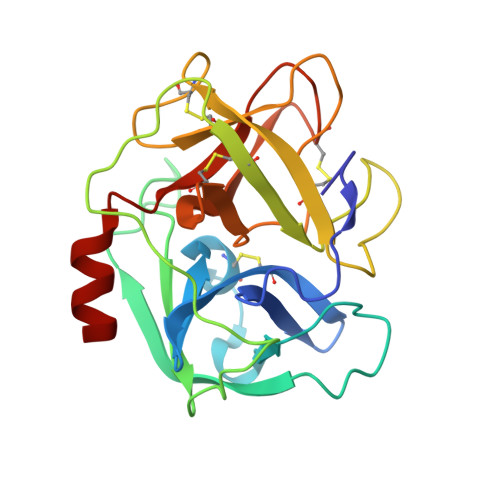
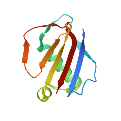
Characterization of two distinct neutrophil serine protease-binding modes within a Staphylococcus aureus innate immune evasion protein family.

Gido, C.D.Herdendorf, T.J.Geisbrecht, B.V.

(2023) J Biol Chem 299: 102969-102969

  • DOI: https://doi.org/10.1016/j.jbc.2023.102969
  • Primary Citation of Related Structures:  
    8D4O, 8D4Q, 8D4S, 8D4U, 8D4V

  • PubMed Abstract: 

    Extracellular adherence protein domain (EAPs) proteins are a class of innate immune evasion proteins secreted by the human pathogen Staphylococcus aureus. EAPs are potent and selective inhibitors of cathepsin-G (CG) and neutrophil elastase (NE), which are the two most abundant neutrophil serine proteases (NSPs). Previous work from our group has shown that the prototypical EAP, EapH1, relies on plasticity within a single inhibitory site to block the activities of CG and NE. However, whether other EAPs follow similar structure-function relationships is unclear. To address this question, we studied the inhibitory properties of the first (Eap1) and second (Eap2) domains of the modular extracellular adherence protein of S. aureus and determined their structures when bound to CG and NE, respectively. We observed that both Eap1 and Eap2 displayed time-dependent inhibition of CG (on the order of 10 -9  M) and of NE (on the order of 10 -10  M). We also found that whereas the structures of Eap1 and Eap2 bound to CG showed an overall inhibitory mode like that seen previously for EapH1, the structures of Eap1 and Eap2 bound to NE revealed a new inhibitory mode involving a distal region of the EAP domain. Using site-directed mutagenesis of Eap1 and Eap2, along with enzyme assays, we confirmed the roles of interfacial residues in NSP inhibition. Taken together, our work demonstrates that EAPs can form structurally divergent complexes with two closely related serine proteases and further suggests that certain EAPs may be capable of inhibiting two NSPs simultaneously.


  • Organizational Affiliation

    Department of Biochemistry & Molecular Biophysics, Kansas State University, Manhattan, Kansas, USA.


Macromolecules
Find similar proteins by:  (by identity cutoff)  |  3D Structure
Entity ID: 1
MoleculeChains Sequence LengthOrganismDetailsImage
Neutrophil elastaseA,
C [auth B]
218Homo sapiensMutation(s): 0 
EC: 3.4.21.37
UniProt & NIH Common Fund Data Resources
Find proteins for P08246 (Homo sapiens)
Explore P08246 
Go to UniProtKB:  P08246
PHAROS:  P08246
GTEx:  ENSG00000197561 
Entity Groups  
Sequence Clusters30% Identity50% Identity70% Identity90% Identity95% Identity100% Identity
UniProt GroupP08246
Sequence Annotations
Expand
  • Reference Sequence
Find similar proteins by:  (by identity cutoff)  |  3D Structure
Entity ID: 2
MoleculeChains Sequence LengthOrganismDetailsImage
Extracellular Adherence ProteinB [auth C],
D
100Staphylococcus aureus subsp. aureus Mu50Mutation(s): 0 
Gene Names: mapSAV1938
UniProt
Find proteins for Q99QS1 (Staphylococcus aureus (strain Mu50 / ATCC 700699))
Explore Q99QS1 
Go to UniProtKB:  Q99QS1
Entity Groups  
Sequence Clusters30% Identity50% Identity70% Identity90% Identity95% Identity100% Identity
UniProt GroupQ99QS1
Sequence Annotations
Expand
  • Reference Sequence
Oligosaccharides

Help

Entity ID: 3
MoleculeChains Length2D Diagram Glycosylation3D Interactions
2-acetamido-2-deoxy-beta-D-glucopyranose-(1-4)-[alpha-L-fucopyranose-(1-6)]2-acetamido-2-deoxy-beta-D-glucopyranose
E, F
3N-Glycosylation
Glycosylation Resources
GlyTouCan:  G21290RB
GlyCosmos:  G21290RB
GlyGen:  G21290RB
Small Molecules
Ligands 1 Unique
IDChains Name / Formula / InChI Key2D Diagram3D Interactions
NAG
Query on NAG

Download Ideal Coordinates CCD File 
G [auth B]2-acetamido-2-deoxy-beta-D-glucopyranose
C8 H15 N O6
OVRNDRQMDRJTHS-FMDGEEDCSA-N
Experimental Data & Validation

Experimental Data

  • Method: X-RAY DIFFRACTION
  • Resolution: 1.90 Å
  • R-Value Free: 0.230 
  • R-Value Work: 0.200 
  • R-Value Observed: 0.201 
  • Space Group: C 1 2 1
Unit Cell:
Length ( Å )Angle ( ˚ )
a = 152.507α = 90
b = 56.009β = 95.05
c = 86.041γ = 90
Software Package:
Software NamePurpose
PHENIXrefinement
HKL-2000data reduction
HKL-2000data scaling
PHASERphasing
PDB_EXTRACTdata extraction

Structure Validation

View Full Validation Report


Ligand Structure Quality Assessment 


Entry History & Funding Information

Deposition Data


Funding OrganizationLocationGrant Number
National Institutes of Health/National Institute of General Medical Sciences (NIH/NIGMS)United States5R35GM140852

Revision History  (Full details and data files)

  • Version 1.0: 2022-12-21
    Type: Initial release
  • Version 1.1: 2023-07-05
    Changes: Database references
  • Version 1.2: 2023-10-25
    Changes: Data collection, Refinement description