8D08

Hallucinated C4 protein assembly HALC4_135


Experimental Data Snapshot

  • Method: X-RAY DIFFRACTION
  • Resolution: 3.30 Å
  • R-Value Free: 0.298 
  • R-Value Work: 0.250 
  • R-Value Observed: 0.253 

wwPDB Validation   3D Report Full Report


This is version 1.2 of the entry. See complete history


Literature

Hallucinating symmetric protein assemblies.

Wicky, B.I.M.Milles, L.F.Courbet, A.Ragotte, R.J.Dauparas, J.Kinfu, E.Tipps, S.Kibler, R.D.Baek, M.DiMaio, F.Li, X.Carter, L.Kang, A.Nguyen, H.Bera, A.K.Baker, D.

(2022) Science 378: 56-61

  • DOI: https://doi.org/10.1126/science.add1964
  • Primary Citation of Related Structures:  
    8D03, 8D04, 8D05, 8D06, 8D07, 8D08, 8D09

  • PubMed Abstract: 

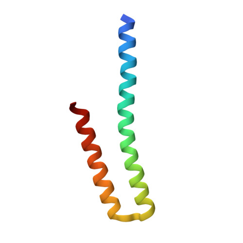
    Deep learning generative approaches provide an opportunity to broadly explore protein structure space beyond the sequences and structures of natural proteins. Here, we use deep network hallucination to generate a wide range of symmetric protein homo-oligomers given only a specification of the number of protomers and the protomer length. Crystal structures of seven designs are very similar to the computational models (median root mean square deviation: 0.6 angstroms), as are three cryo-electron microscopy structures of giant 10-nanometer rings with up to 1550 residues and C 33 symmetry; all differ considerably from previously solved structures. Our results highlight the rich diversity of new protein structures that can be generated using deep learning and pave the way for the design of increasingly complex components for nanomachines and biomaterials.


  • Organizational Affiliation

    Department of Biochemistry, University of Washington, Seattle, WA, USA.


Macromolecules
Find similar proteins by:  (by identity cutoff)  |  3D Structure
Entity ID: 1
MoleculeChains Sequence LengthOrganismDetailsImage
HALC4_135
A, B, C, D
70synthetic constructMutation(s): 0 
Entity Groups  
Sequence Clusters30% Identity50% Identity70% Identity90% Identity95% Identity100% Identity
Sequence Annotations
Expand
  • Reference Sequence
Experimental Data & Validation

Experimental Data

  • Method: X-RAY DIFFRACTION
  • Resolution: 3.30 Å
  • R-Value Free: 0.298 
  • R-Value Work: 0.250 
  • R-Value Observed: 0.253 
  • Space Group: P 41 21 2
Unit Cell:
Length ( Å )Angle ( ˚ )
a = 35.867α = 90
b = 35.867β = 90
c = 437.996γ = 90
Software Package:
Software NamePurpose
PHENIXrefinement
XDSdata reduction
XSCALEdata scaling
PHASERphasing

Structure Validation

View Full Validation Report


Entry History & Funding Information

Deposition Data


Funding OrganizationLocationGrant Number
Howard Hughes Medical Institute (HHMI)United States--

Revision History  (Full details and data files)

  • Version 1.0: 2022-09-28
    Type: Initial release
  • Version 1.1: 2022-10-19
    Changes: Database references
  • Version 1.2: 2024-04-03
    Changes: Data collection, Refinement description