8CZG

Human BAK in complex with the dF3 peptide


Experimental Data Snapshot

  • Method: X-RAY DIFFRACTION
  • Resolution: 1.99 Å
  • R-Value Free: 0.267 
  • R-Value Work: 0.228 
  • R-Value Observed: 0.230 

wwPDB Validation   3D Report Full Report


This is version 1.3 of the entry. See complete history


Literature

Peptides from human BNIP5 and PXT1 and non-native binders of pro-apoptotic BAK can directly activate or inhibit BAK-mediated membrane permeabilization.

Aguilar, F.Yu, S.Grant, R.A.Swanson, S.Ghose, D.Su, B.G.Sarosiek, K.A.Keating, A.E.

(2023) Structure 31: 265-281.e7

  • DOI: https://doi.org/10.1016/j.str.2023.01.001
  • Primary Citation of Related Structures:  
    8CZF, 8CZG, 8CZH

  • PubMed Abstract: 

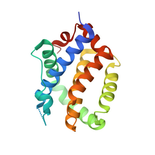
    Apoptosis is important for development and tissue homeostasis, and its dysregulation can lead to diseases, including cancer. As an apoptotic effector, BAK undergoes conformational changes that promote mitochondrial outer membrane disruption, leading to cell death. This is termed "activation" and can be induced by peptides from the human proteins BID, BIM, and PUMA. To identify additional peptides that can regulate BAK, we used computational protein design, yeast surface display screening, and structure-based energy scoring to identify 10 diverse new binders. We discovered peptides from the human proteins BNIP5 and PXT1 and three non-native peptides that activate BAK in liposome assays and induce cytochrome c release from mitochondria. Crystal structures and binding studies reveal a high degree of similarity among peptide activators and inhibitors, ruling out a simple function-determining property. Our results shed light on the vast peptide sequence space that can regulate BAK function and will guide the design of BAK-modulating tools and therapeutics.


  • Organizational Affiliation

    Department of Biology, Massachusetts Institute of Technology, Cambridge, MA, USA.


Macromolecules
Find similar proteins by:  (by identity cutoff)  |  3D Structure
Entity ID: 1
MoleculeChains Sequence LengthOrganismDetailsImage
Bcl-2 homologous antagonist/killer
A, B, C, D
170Homo sapiensMutation(s): 1 
Gene Names: BAK1BAKBCL2L7CDN1
UniProt & NIH Common Fund Data Resources
Find proteins for Q16611 (Homo sapiens)
Explore Q16611 
Go to UniProtKB:  Q16611
PHAROS:  Q16611
GTEx:  ENSG00000030110 
Entity Groups  
Sequence Clusters30% Identity50% Identity70% Identity90% Identity95% Identity100% Identity
UniProt GroupQ16611
Sequence Annotations
Expand
  • Reference Sequence

Find similar proteins by:  Sequence   |   3D Structure  

Entity ID: 2
MoleculeChains Sequence LengthOrganismDetailsImage
dF3 peptide
E, F, G, H
25synthetic constructMutation(s): 0 
Entity Groups  
Sequence Clusters30% Identity50% Identity70% Identity90% Identity95% Identity100% Identity
Sequence Annotations
Expand
  • Reference Sequence
Experimental Data & Validation

Experimental Data

  • Method: X-RAY DIFFRACTION
  • Resolution: 1.99 Å
  • R-Value Free: 0.267 
  • R-Value Work: 0.228 
  • R-Value Observed: 0.230 
  • Space Group: P 1 21 1
Unit Cell:
Length ( Å )Angle ( ˚ )
a = 48.29α = 90
b = 65.26β = 102.12
c = 111.57γ = 90
Software Package:
Software NamePurpose
PHENIXrefinement
XDSdata reduction
XSCALEdata scaling
PHASERphasing
PDB_EXTRACTdata extraction

Structure Validation

View Full Validation Report


Entry History & Funding Information

Deposition Data


Funding OrganizationLocationGrant Number
National Institutes of Health/National Institute of General Medical Sciences (NIH/NIGMS)United StatesR01GM110048

Revision History  (Full details and data files)

  • Version 1.0: 2023-01-11
    Type: Initial release
  • Version 1.1: 2023-02-08
    Changes: Database references
  • Version 1.2: 2023-03-15
    Changes: Database references
  • Version 1.3: 2023-10-25
    Changes: Data collection, Refinement description