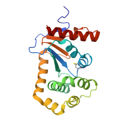
8CXD

Crystal Structure of EcDsbA in a complex with phenylmethanol


Experimental Data Snapshot

  • Method: X-RAY DIFFRACTION
  • Resolution: 1.80 Å
  • R-Value Free: 0.200 
  • R-Value Work: 0.175 
  • R-Value Observed: 0.176 

wwPDB Validation   3D Report Full Report


Ligand Structure Quality Assessment 


This is version 1.2 of the entry. See complete history


Literature

Fragment screening libraries for the identification of protein hot spots and their minimal binding pharmacophores.

Whitehouse, R.L.Alwan, W.S.Ilyichova, O.V.Taylor, A.J.Chandrashekaran, I.R.Mohanty, B.Doak, B.C.Scanlon, M.J.

(2023) RSC Med Chem 14: 135-143

  • DOI: https://doi.org/10.1039/d2md00253a
  • Primary Citation of Related Structures:  
    8CXD, 8CXE, 8CZM, 8CZN, 8D10, 8D11, 8D12, 8DG0, 8DG1, 8DG2

  • PubMed Abstract: 

    Fragment-based drug design relies heavily on structural information for the elaboration and optimisation of hits. The ability to identify neighbouring binding hot spots, energetically favourable interactions and conserved binding motifs in protein structures through X-ray crystallography can inform the evolution of fragments into lead-like compounds through structure-based design. The composition of fragment libraries can be designed and curated to fit this purpose and herein, we describe and compare screening libraries containing compounds comprising between 2 and 18 heavy atoms. We evaluate the properties of the compounds in these libraries and assess their ability to probe protein surfaces for binding hot spots.


  • Organizational Affiliation

    Medicinal Chemistry, Monash Institute of Pharmaceutical Sciences, Monash University Parkville VIC 3052 Australia martin.scanlon@monash.edu.


Macromolecules
Find similar proteins by:  (by identity cutoff)  |  3D Structure
Entity ID: 1
MoleculeChains Sequence LengthOrganismDetailsImage
Thiol:disulfide interchange protein DsbA
A, B
189Escherichia coli K-12Mutation(s): 0 
Gene Names: dsbAdsfppfAb3860JW3832
UniProt
Find proteins for P0AEG4 (Escherichia coli (strain K12))
Explore P0AEG4 
Go to UniProtKB:  P0AEG4
Entity Groups  
Sequence Clusters30% Identity50% Identity70% Identity90% Identity95% Identity100% Identity
UniProt GroupP0AEG4
Sequence Annotations
Expand
  • Reference Sequence
Experimental Data & Validation

Experimental Data

  • Method: X-RAY DIFFRACTION
  • Resolution: 1.80 Å
  • R-Value Free: 0.200 
  • R-Value Work: 0.175 
  • R-Value Observed: 0.176 
  • Space Group: C 1 2 1
Unit Cell:
Length ( Å )Angle ( ˚ )
a = 117.725α = 90
b = 62.577β = 126.347
c = 74.722γ = 90
Software Package:
Software NamePurpose
PHENIXrefinement
XDSdata reduction
Aimlessdata scaling
PHASERphasing

Structure Validation

View Full Validation Report


Ligand Structure Quality Assessment 


Entry History & Funding Information

Deposition Data


Funding OrganizationLocationGrant Number
Australian Research Council (ARC)AustraliaIC180100021
Australian Research Council (ARC)AustraliaDP200100796

Revision History  (Full details and data files)

  • Version 1.0: 2022-12-07
    Type: Initial release
  • Version 1.1: 2023-02-22
    Changes: Database references
  • Version 1.2: 2023-10-25
    Changes: Data collection, Refinement description