


Crystal Structure of dTDP-4-dehydrorhamnose reductase from Klebsiella pneumoniae with bound NADP
Bolejack, M.J., Abendroth, J., Lorimer, D.D., Horanyi, P.S., Edwards, T.E.To be published.
Experimental Data Snapshot
Starting Model: experimental
View more details
Entity ID: 1 | |||||
|---|---|---|---|---|---|
| Molecule | Chains | Sequence Length | Organism | Details | Image |
| dTDP-4-dehydrorhamnose reductase | 304 | Klebsiella pneumoniae | Mutation(s): 0 Gene Names: rmlD, rfbD, rfbD_1, rfbD_2, A7B01_07910, C3F39_24350, DM23092/04_00022, GNE24_11460, SAMEA3649733_05275, SAMEA3727643_01606... EC: 1.1.1.133 | 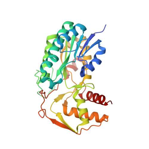 | |
UniProt | |||||
Entity Groups | |||||
| Sequence Clusters | 30% Identity50% Identity70% Identity90% Identity95% Identity100% Identity | ||||
| UniProt Group | C9K1F1 | ||||
Sequence AnnotationsExpand | |||||
Reference Sequence | |||||
| Ligands 4 Unique | |||||
|---|---|---|---|---|---|
| ID | Chains | Name / Formula / InChI Key | 2D Diagram | 3D Interactions | |
| NAP (Subject of Investigation/LOI) Download:Ideal Coordinates CCD File | E [auth A], J [auth B], N [auth C], Q [auth D] | NADP NICOTINAMIDE-ADENINE-DINUCLEOTIDE PHOSPHATE C21 H28 N7 O17 P3 XJLXINKUBYWONI-NNYOXOHSSA-N |  | ||
| GOL Download:Ideal Coordinates CCD File | G [auth A], I [auth B], P [auth C], S [auth D] | GLYCEROL C3 H8 O3 PEDCQBHIVMGVHV-UHFFFAOYSA-N |  | ||
| ACT Download:Ideal Coordinates CCD File | F [auth A], K [auth B], O [auth C], R [auth D] | ACETATE ION C2 H3 O2 QTBSBXVTEAMEQO-UHFFFAOYSA-M |  | ||
| NA Download:Ideal Coordinates CCD File | H [auth A], L [auth B], M [auth B], T [auth D] | SODIUM ION Na FKNQFGJONOIPTF-UHFFFAOYSA-N |  | ||
| Length ( Å ) | Angle ( ˚ ) |
|---|---|
| a = 71.55 | α = 89.96 |
| b = 71.69 | β = 66.58 |
| c = 81.23 | γ = 83.08 |
| Software Name | Purpose |
|---|---|
| XDS | data reduction |
| XSCALE | data scaling |
| PHENIX | refinement |
| PDB_EXTRACT | data extraction |
| MoRDa | phasing |
| Funding Organization | Location | Grant Number |
|---|---|---|
| National Institutes of Health/National Institute Of Allergy and Infectious Diseases (NIH/NIAID) | United States | -- |


